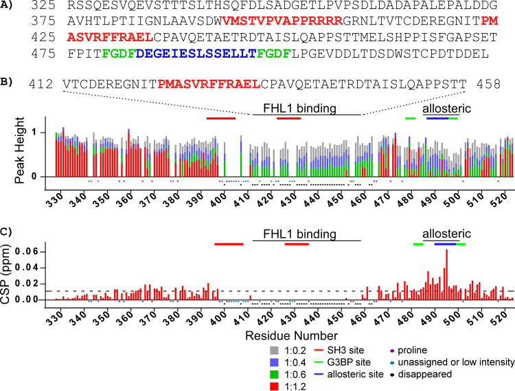
FIG 8.
Changes of the CHIKV HVD amide resonances in 1H-15N BEST-TROSY spectra upon titration with FHL1. (A) The amino acid sequence of CHIKV nsP3 HVD, which was used in the experiments. The previously identified CD2AP- and G3BP-binding and allosteric sites are indicated in red, green, and blue, respectively. Both the changes in normalized intensities (peak height) and chemical shift perturbations (CSPs) are shown in panels B and C, respectively. In panel C, the dashed line shows the level of 2 standard deviations of variation of CS during titration. Ratios of titrations used are shown below panel C. Prolines, unassigned amino acids, low intensities, or cross-peaks disappearing during titration are indicated below panel C. Marked above in red solid lines are the CD2AP-binding sites, in blue are the proposed CD2AP-specific allosteric site, and in green are the G3BP-binding sites. They are marked correspondingly to the amino acid sequence in panel A.

