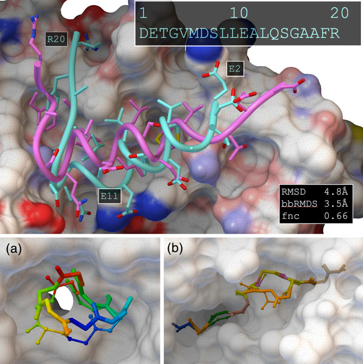
FIGURE 7.

Top panel: best docking solution (cyan) and crystal structure (magenta) of a linear peptide that forms a self‐inhibitory switch in formin mDia1 (PDB entry 2f31). Starting from the sequence, ADCP folds and places the peptide into the receptor. Sidechain‐receptor interactions are predicted well, except for E2, E11, and R20 that find polar patches. Bottom panel: backbone of docked cyclic peptides (ball‐and‐stick) and crystal structure (licorice). (a) Top ranked docked pose for a ubiquitin ligase substrate adaptor protein with an engineered cyclized peptide from one of its targets (PDB entry 3zgc). Residues are colored blue to red to show correct registration. (b) Docking result for an internally disulfide‐linked peptide from Epstein Barr Virus being displayed by MHC (PDB entry 5grd). Residues are color‐coded by residue type and the side chains of the two cysteines show the created disulfide bond
