Figure 2.

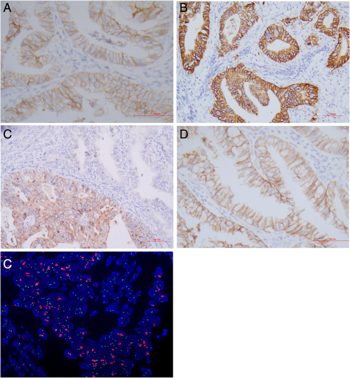
Detection of HER2 in cervical adenocarcinoma. (A, B) Representative areas of HER2 expression 2+, and 3+, respectively. (C) Heterogeneous HER2 staining. (D) Incomplete membranous (‘U’‐shaped) immunoreactivity. (E) HER2 amplification by FISH. Immunohistochemistry: A–D; FISH: E. Original magnifications: A–C ×200; D, E ×400.
