Abstract
Background:
Studies show that fluoride (F) and nano-hydroxyapatite (nano-HA) would result in remineralization of white spot lesions (WSLs), which are among the most prevalent consequences of fixed orthodontic treatment. The present study evaluates and compares the clinical effects of an Iranian toothpaste containing nano-HA with F-containing one on early enamel lesions.
Materials and Methods:
In this randomized clinical trial study, 50 patients who had received fixed orthodontic treatment were recruited immediately after debonding. Three photographs, including frontal, lateral right and left views of occlusion, were obtained. Moreover, surfaces with WSLs were recorded using DIAGNOdent. Plaque index of each patient determined using disclosing agents. At first visit, each patient was asked to select one type of toothpaste (nano-HA containing vs. F containing named A or B), randomly and were instructed how to brush their teeth (25 patients in each group). Examination was done at 1, 3, and 6 months' intervals. Finally, photographs were analyzed by Digimizer (V5) software, and the lesion extent was recorded in pixels. SAS 9.4 was used to analyze data and was set at 0.05.
Results:
According to data, lesion extent showed a significant decrease (P < 0.001). At baseline, the difference between the two groups regarding the lesion extent was 268 pixels while it dropped to 89 pixels after 6 months. DIAGNOdent results showed that at baseline, fluorescence difference was 0.3 while it reached the number of 0.8 after 6 months, indicating the outperformance of nano-HA containing toothpaste.
Conclusion:
The Iranian nano-HA containing toothpaste performed better than F-containing one in terms of the amount of remineralization and diminishing the lesion extent.
Key Words: Fluoride, nano-hydroxyapatite, toothpaste, white spot lesion
INTRODUCTION
Following orthodontic treatment, the oral environment undergoes several changes and white spot lesions (WSLs) due to plaque accumulation and poor oral hygiene. If remain untreated, restorative treatments could be inevitable. Moreover, they adversely affect patients' appearance.[1,2]
During orthodontic treatment, acidogenic bacteria such as Streptococcus mutans increase in numbers which lead to a reduction in oral PH and development of decalcification and early enamel lesions.[3] Management and treatment of such lesions are one of the major challenges that orthodontists confront. The primary and feasible way of prevention is oral hygiene instructions to diminish plaque accumulation and frequency of lesions.[4] Other strategies include applying toothpastes containing nanohydroxyapatite (nano-HA), fluoride (F), gels containing F, restorative, and preventive treatments among which, utilizing toothpaste is usually considered to be more accessible and convenient for patients.[5]
Toothpastes containing nano-HA provide dentin tubules and enamel rods with HA particles, which result in their remineralization. This procedure inhibits the lesion progress.[6,7,8] Recently, a new generation of such toothpastes has been developed, which contains HA particles of nano size. These toothpastes perform better than F-containing ones in terms of remineralizing teeth.[9]
Nano-HA has a great infinity to infiltrate into enamel rods due to its small size and has a high potential for restoring lesions. Japanese have incorporated this element in the toothpaste since 1980.[9,10] Nano-HA containing toothpastes have been converted to one of the most applicable products to attack WSLs due to their function in remineralization. Huang et al. found that the concentration of 10% nano-HA would have the potential to remineralize initial enamel defects.[11] In addition, Tschoppe et al. revealed that toothpaste containing nano-HA has a higher remineralizing effect compared to amine fluoride toothpaste on dentin and enamel lesions.[12] In Iran, only foreign brands exist commercially; thus, many people have difficulty affording such toothpastes. In the present study, the efficacy and effectiveness of one of the newly developed Iranian toothpastes containing nano-HA are evaluated in vivo.
MATERIALS AND METHODS
Stages of developing toothpaste
First, the gelling agent was dispensed in protected water containing sweeteners and other carriers. Then, the abrasive agent was added and beaten using a digital laboratory stirrer (IKA EUROSTAR Power Control-Visc Stirrer-Germany). The preservative was added, and finally, a laboratory-scale homogenizing system (STEPHAN UMC 5-Germany) was used to make the product uniform. The product then evaluated its physical stability in refrigerator, room and 40°C temperatures. Mechanical activation as well as Sol-Gel method is usually applied to produce nano-HA. We selected the Sol-Gel procedure. Then, nanoparticles of 4.5% concentration were added to an ordinary toothpaste.
Sol–Gel method
NO3.4H2O. CA and P2O5 were used as basic elements and were mixed using a mechanical mixer until a stable and clear Sol was made. Consolidation and polymerization reactions were completed in environment after 24 h, and the Gel was constructed. Then, it was dried at 600°C.
Method
In this randomized clinical trial (IRCT20180928041162N1), 80 patients who had undergone orthodontic treatment in the Orthodontics Department of Shahid Beheshti University of Medical Sciences and had WSLs on facial surfaces of their teeth were included immediately after debonding. Thirty patients were excluded due to un-cooperation and poor oral hygiene. Finally, 50 patients (34% males and 66% females) with the age range of 10–35 years were included in the study and allocated to the groups using the stratified permutation blocks to match the confounding variables and ensure about equality of sample sizes in the groups.
This sample size was calculated based on type-one error of 0.05 and type-two error of 0.2 (power = 80%) and effect size of 0.6 according to previous studies using an appropriate statistical formula.
Inclusion criteria were as follows:
Samples were recruited from patients who used Hawley retainers
Patients having WSL on the buccal surface of at least one tooth
Patient's consent for participating in the study
Good oral hygiene
Patient cooperation in using the allocated toothpaste according to the instructions.
At first, study procedures were explained to them. In the primary examination, each patient was given a toothpaste of type A (nano-HA 6.7%) or B (ordinary F containing toothpaste) randomly (toothpastes were coded A or B, and they were offered to patients in succession) that the practitioner was also blind (double-blind study). Demographic information (name, family name, address, and contact number) were collected. A group of three photographs was obtained and evaluated using DIAGNOdent (Kavo pen DIAGNOdent 2190, Germany). Plaque index of each patient was recorded by disclosing pills also, the degree of WSLs was determined, and all procedures were repeated in 1–3 and 6 months' intervals. Data registered were as follows:
White spot lesion degree
The International Caries Detection and Assessment System index, which is related to enamel lesions, was used to determine the degree of lesions. The practitioner clarified the degree of lesions by using the given table and observation of teeth. Lesions of Grade 2 and 3 were just included in the present study.
DIAGNOdent examination and determining fluorescence value
In order to determine fluorescence value Kavodiagnodent (Kavo pen DIAGNOdent 2190, Germany) was used which have two tips to examine facial and proximal surfaces. We used the facial type to evaluate the early enamel lesions on buccal surfaces of teeth. At first, teeth were dried then, DIAGNOdent was calibrated with the nearest tooth to the tooth containing lesion. The test tooth should lack any kind of stain and caries as to be the reference. Then, the tooth with lesion was examined using DIAGNOdent under daylight, and it was repeated after 1 min. The higher value was recorded (the differences in obtained values in various stages reveal the changes that occurred in enamel fluorescence).
Photography and its analysis
Patients sat on a chair in natural head Position while their lips and cheeks were retracted. Photographs were taken from right, left, and frontal views. A Canon 600d camera (Canon, Japan) and a 60 mm lens were used. Diaphragm was sat up at 32 and shutter pace at 320. Lens focus was on canine and incisors in frontal view while in lateral one, the focus was on premolars and molars. Camera angulation was perpendicular to central incisors in frontal views while in lateral views, it was sat perpendicular to canines to obtain figures of all molars. In order to standardize and analyze photographs, first, their sizes were unified (1000 × 3000 pixels) by Photoshop software Adobe Inc, US, and then the extent of lesion was measured by the number of relevant pixels and recorded. Finally, the plaque index was evaluated using disclosing pills and calculated according to the formula:
The number of colored surfaces
The number of all surfaces
The method of Bass brushing was instructed, and patients were said to use the toothpaste twice a day (mornings and nights). Examinations were repeated at 1-, 3-, and 6-month intervals, and at each stage, all methods were explained again, and patients were given the toothpaste which was applied before.
Descriptive statistics, including the mean, percentage, standard deviation along with tables and figures, were used. In inferential statistics, according to the 3-level structure of the data, multilevel-linear mixed model which is a specific type of random-effect model was used for dependent variables such as plaque index, pixels of lesion extent, and fluorescence number. Type-one error was set at 0.05, and analysis was done with SAS version 9.4 SAS (Institute Inc, US). The baseline data were entered into the model, and their effects were adjusted, so they did not have a confounder role.
RESULTS
Demographic data
One hundred and seventy-three teeth were studied (59 males [34%] and 114 females [66%]). Ninety-six teeth (55%) were belonged to patients younger than 20, 69 teeth (40%) to 20–30 years old patients and 8 samples (5%) were evaluated of patients older than 30.
The evaluation of dependent variables
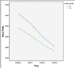
Lesion extent divided by the studied toothpaste: At all time-intervals, the extent of lesion at baseline showed no significant difference between two groups (P = 0.736), but as the time passed, the extent of lesion significantly decreased (P < 0.0001). Moreover, the effect of toothpaste was significant; meanwhile, both groups showed an approximate difference of 268 pixels in lesion extent at baseline while following 6 months' application of toothpastes, the difference decreased to 82 pixels, which represents the outperformance of nano-HA containing toothpaste [Graph 1].
Graph 1.

Average changes in lesion size in two types of toothpaste.
Variables, including age and gender, have shown no significant effect on this trend (P = 0.962 and P = 0.824, respectively).
DIAGNOdent evaluation
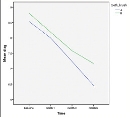
Graph 2 demonstrates the results of multilevel model analysis of DIAGNOdent data. The mean value of DIAGNOdent data at baseline was not significantly different between the two groups (P = 0.878), but the reduction in this value was significant during the time intervals (P = 0.009).
Graph 2.

Average variation of DIAGNOdent numbers in 2 types of toothpaste over time.
Furthermore, the effect of the type of toothpaste on DIAGNOdent data was significant, so that the fluorescence difference was recorded 0.3 units at baseline while after 6 months, it reached 0.8 units. According to Graph 2, nano-HA containing toothpaste showed a greater decrease. Gender (P = 0.473) and age (P = 0.959) did not affect fluorescence, significantly.
Plaque index
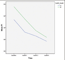
Table 1 provides data about plaque indexes of studied teeth. According to the results of multilevel model related to plaque index, the amount of accumulated plaque has undergone a significant decrease in either group after 6 months, and this reduction was significantly different between two groups (P < 0.001). At baseline, there was a difference of 6 while it reduced to 2 following 6 months, although it was not clinically significant [Graph 3].
Table 1.
Statistical indicators of plaque index in two types of toothpaste during time intervals
| Time | Toothpaste type | Mean | n | SD |
|---|---|---|---|---|
| Base | (A) Nano type | 48.44 | 77 | 0.992 |
| (B) F type | 53.96 | 96 | 1.125 | |
| 1 month | (A) Nano type | 43.06 | 77 | 0.703 |
| (B) F type | 48.65 | 96 | 1.061 | |
| 3 months | (A) Nano type | 41.43 | 77 | 0.836 |
| (B) F type | 43.74 | 96 | 0.839 | |
| 6 months | (A) Nano type | 39.16 | 77 | 0.808 |
| (B) F type | 40.83 | 96 | 0.717 |
F: Fluoride; SD: Standard deviation
Graph 3.

Average variation of plaque index in 2 types of toothpaste over time.
DISCUSSION
According to the recent advances in technology, many problems that encountered people have been solved among which is dental caries that always have been considered as a reason of tooth loss. Dental caries and associated lesions such as WSL are one of the most frequent side effects of fixed orthodontic treatment, and one of the greatest challenges that orthodontists face is how to prevent and diminish such lesions.[3] Several materials have been introduced to solve the problem such as the application of nano-HA containing toothpastes, fluoride, microabrasion, and toothpastes containing amorphous Ca/P which contain advantages and disadvantages. Recent improvements in nanoscience and its integration into dental material science have resulted in the production of new materials with improved functionality such as nano-HA.[10]
According to its remineralization activity, we aimed to compare the remineralizing effect of toothpaste containing nano-HA made in Iran with a fluoride-containing toothpaste.
Haghgoo et al. provided 18 enamel blocks from human-impacted third molars in an experimental in vitro study. They targeted at comparing solutions of 10% nano-HA with water and utilized Vicker's test to record the samples' microhardness. They used carbonated drinks to simulate lesions. Similar to our findings, they concluded that nano-HA made enamel blocks much harder.[14] Although in another study conducted by the same author, they compared the remineralizing effect of a toothpaste containing different concentrations of nano-HA on incipient caries. Concentrations of 0%, 0.5%, 1%, 2%, and 5% wt of nano-HA were added to a solution of distilled water and a toothpaste. Results showed that nano-HA at all concentrations increased the microhardness of demineralized teeth; however, it was not statistically significant.[13]
Tschoppe et al., in an experimental study on 70 bovine enamel and 80 dentin blocks, evaluated the effect of different concentrations of nano-HA and F on remineralizing the enamel lesions. They found no significant difference among various concentrations of nano-HA while it outperformed F. Their results are consistent to ours.[12]
Yuan et al. used dentin of 24 premolar and 24 molar teeth to produce samples and compared the toothpaste containing 3% nano-HA with an ordinary one. They exposed dentinal tubules to make lesions, and applied method was Energy-Dispersive X-ray Spectroscopy (to measure the mineral content). They also reported that the ability of tubular obstruction and remineralization of nano-HA is significant.[15]
Consistently, Ebadifar et al. conducted a study on 80 enamel blocks, which were demineralized using phosphoric acid. Wickers' test was used, and they concluded that nano-HA containing toothpaste overweigh F containing one.[16]
Conversely, Najibfard et al. used 4 enamel blocks of third molars to compare solutions of 5%, 10% nano-HA, and 10% nano-HA/NaF 1100 ppm. They found no significant difference among various concentrations of nano-HA in remineralizing potential. It might be explained by the different methods used for measuring remineralization. They applied microradiography which does not have the appropriate accuracy in recording the amount of remineralization.[9]
Jeong et al. studied on 22 human enamel blocks with the aim of comparing pure nano-HA solution with a solution containing nano-HA/NaF 0.65%. Wickers' microhardness test was used, and similarly, they reported that nano-HA containing toothpaste performed better.[8]
In the present study, we used two indexes of lesion extent and fluorescence degree (which has a direct correlation to the amount of remineralization) as to evaluate the effect of toothpaste on WSLs. Photographs were provided to compare the lesion extent at baseline, 1-, 3-, and 6-month intervals which showed that nano-HA containing toothpaste resulted in a significant reduction in its extent after 6 months. The reduction trend followed from 1 to 6 months predicts that continuous application of toothpaste might result in a greater decrease by passing time.
We used DIAGNOdent to measure the fluorescence which found that the final reduction in its value occurred in the nano-HA group. Similarly, Bahrololoomi et al. showed that the fluorescence value would decrease by improvement in remineralization.[17]
In the present study, F group also showed some levels of decrease in lesion extent and fluorescence which is consistent with the findings of Willmot who compared low F and high F toothpastes in remineralizing enamel lesions following orthodontic treatment. They reported that F can reduce the size of these lesions.[18] Moreover, Korkmaz and Yagci also reported that F-releasing agents can prevent WSLs in patients treated with full coverage of rapid maxillary expanders.[19]
Our study has some superiorities over previous ones as we evaluated the lesion extent as well as the degree of remineralization, while previous studies did not examine the clinical effects of nano-HA on lesion extents. In addition, it was the first time that a national toothpaste was tested in vivo.
The plaque index was also recorded as a measure of plaque control. Six months' follow-up showed a decrease in this value in both groups, which reveals that either group had good oral hygiene.
Although one of the most important limitations of the study was to keep the patients, cooperative during the 6-month follow-up.
CONCLUSION
Nano-HA containing toothpaste performs better than F containing toothpaste in terms of remineralization
Age and gender do not affect the remineralizing potential
During 6-month period, both agents resulted in improved remineralization, while nano-HA performs better
Nano-HA containing toothpaste is an appropriate method of controlling early enamel lesions.
Financial support and sponsorship
This study was financially supported by both Goltash Co. and Dentofacial Deformities Research Center at Shahid Beheshti University of Medical Sciences.
Conflicts of interest
The authors of this manuscript declare that they have no conflicts of interest, real or perceived, financial or non-financial in this article.
Acknowledgment
The authors would like to thank the patients who participated in this survey for their cooperation and completing informed consent.
REFERENCES
- 1.Kronenberg O, Lussi A, Ruf S. Preventive effect of ozone on the development of white spot lesions during multibracket appliance therapy. Angle Orthod. 2009;79:64–9. doi: 10.2319/100107-468.1. [DOI] [PubMed] [Google Scholar]
- 2.Sudjalim TR, Woods MG, Manton DJ. Prevention of white spot lesions in orthodontic practice: A contemporary review. Aust Dent J. 2006;51:284–9. doi: 10.1111/j.1834-7819.2006.tb00445.x. [DOI] [PubMed] [Google Scholar]
- 3.Bishara SE, Ostby AW. White spot lesions: Formation, prevention, and treatment. Semin Orthod. 2008;14:174–82. [Google Scholar]
- 4.Andersson A, Sköld-Larsson K, Hallgren A, Petersson LG, Twetman S. Effect of a dental cream containing amorphous cream phosphate complexes on white spot lesion regression assessed by laser fluorescence. Oral Health Prev Dent. 2007;5:229–33. [PubMed] [Google Scholar]
- 5.Cosma LL, Şuhani RD, Mesaroş A, Badea ME. Current treatment modalities of orthodontically induced white spot lesions and their outcome a literature review. Med Pharm Rep. 2019;92:25–30. doi: 10.15386/cjmed-1090. [DOI] [PMC free article] [PubMed] [Google Scholar]
- 6.Huang S, Gao S, Cheng L, Yu H. Combined effects of nano-hydroxyapatite and galla chinensis on remineralisation of initial enamel lesion in vitro. J Dent. 2010;38:811–9. doi: 10.1016/j.jdent.2010.06.013. [DOI] [PubMed] [Google Scholar]
- 7.Esteves-Oliveira M, Santos NM, Meyer-Lueckel H, Wierichs RJ, Rodrigues JA. Caries-preventive effect of anti-erosive and nano-hydroxyapatite-containing toothpastes in vitro. Clin Oral Investig. 2017;21:291–300. doi: 10.1007/s00784-016-1789-0. [DOI] [PubMed] [Google Scholar]
- 8.Jeong S, Jang S, Kim KN, Kwon H, Park YD, Kim B. Remineralization potential of new toothpaste containing nano-hydroxyapatite. Key Eng Mater. 2006;309:537–40. [Google Scholar]
- 9.Najibfard K, Ramalingam K, Chedjieu I, Amaechi BT. Remineralization of early caries by a nano-hydroxyapatite dentifrice. J Clin Dent. 2011;22:139–43. [PubMed] [Google Scholar]
- 10.Shungin D, Olsson AI, Persson M. Orthodontic treatment-related white spot lesions: A 14-year prospective quantitative follow-up, including bonding material assessment. Am J Orthod Dentofacial Orthop. 2010;138:136.e1–8. doi: 10.1016/j.ajodo.2009.05.020. [DOI] [PubMed] [Google Scholar]
- 11.Huang SB, Gao SS, Yu HY. Effect of nano-hydroxyapatite concentration on remineralization of initial enamel lesion in vitro. Biomed Mater. 2009;4:034104. doi: 10.1088/1748-6041/4/3/034104. [DOI] [PubMed] [Google Scholar]
- 12.Tschoppe P, Zandim DL, Martus P, Kielbassa AM. Enamel and dentine remineralization by nano-hydroxyapatite toothpastes. J Dent. 2011;39:430–7. doi: 10.1016/j.jdent.2011.03.008. [DOI] [PubMed] [Google Scholar]
- 13.Haghgoo R, Abbasi F, Rezvani MB. Evaluation of the effect of nanohydroxyapatite on erosive lesions of the enamel of permanent teeth following exposure to soft beer in vitro. Sci Res Essays. 2011;6:5933–6. [Google Scholar]
- 14.Haghgoo R, Rezvani MR, Haghgoo HR, Ameli N, Zeinabadi MS. Evaluation of Iranian toothpaste containing different concentrations of nano-hydroxyapatite on the remineralization of incipient carious lesions: In vitro. J Dent Med. 2015;27:254–8. [Google Scholar]
- 15.Yuan P, Shen X, Liu J, Hou Y, Zhu M, Huang J, et al. Effects of dentifrice containing hydroxyapatite on dentinal tubule occlusion and aqueous hexavalent chromium cations sorption: A preliminary study. PLoS One. 2012;7:e45283. doi: 10.1371/journal.pone.0045283. [DOI] [PMC free article] [PubMed] [Google Scholar]
- 16.Ebadifar A, Nomani M, Fatemi SA. Effect of nano-hydroxyapatite toothpaste on microhardness ofartificial carious lesions created on extracted teeth. J Dent Res Dent Clin Dent Prospects. 2017;11:14–7. doi: 10.15171/joddd.2017.003. [DOI] [PMC free article] [PubMed] [Google Scholar]
- 17.Bahrololoomi Z, Musavi SA, Kabudan M. In vitro evaluation of the efficacy of laser fluorescence (DIAGNOdent) to detect demineralization and remineralization of smooth enamel lesions. J Conserv Dent. 2013;16:362–6. doi: 10.4103/0972-0707.114360. [DOI] [PMC free article] [PubMed] [Google Scholar]
- 18.Willmot DR. White lesions after orthodontic treatment: Does low fluoride make a difference? J Orthod. 2004;31:235–42. doi: 10.1179/146531204225022443. [DOI] [PubMed] [Google Scholar]
- 19.Korkmaz YN, Yagci A. Comparing the effects of three different fluoride-releasing agents on white spot lesion prevention in patients treated with full coverage rapid maxillary expanders. Clin Oral Investig. 2019;23:3275–85. doi: 10.1007/s00784-018-2749-7. [DOI] [PubMed] [Google Scholar]


