Figure 7.

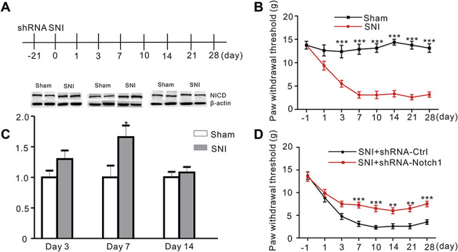
Microinjections of the Notch1 shRNA into the ACC inhibited mechanical allodynia induced by SNI in rats. (A) Schematic of ACC microinjections and behavior experiments. (B) Mechanical withdrawal thresholds were measured in Sham and SNI rats before and 1, 3, 7, 10, 14, 21, and 28 days after surgery. Results are presented as mean ± SME (F = 11.19, P < 0.0001***, n = 8 animals per group, two-way ANOVA followed by the Bonferroni post hoc test, black: Sham, red: SNI). (C) Western blot showing increased NICD levels in the ACC (day 7 after surgery) in the SNI group and had no difference compared to Sham group at day 3 and 14. Bottom panel, quantification of the intensity of the NICD bands (P = 0.0282, Student t-test, n = 8 animals per group). (D) Attenuation of SNI-induced mechanical allodynia by microinjections of shRNA-Notch1 into the ACC (F = 2.831, P = 0.0094, n = 8 animals per group, two-way ANOVA followed by the Bonferroni post hoc test, black: SNI + shRNA-Ctrl, red: SNI + shRNA-Notch1). ACC, anterior cingulate cortex; ANOVA, analysis of variance; NICD, Notch intracellular domain; shRNA, short hairpin RNA; SNI, spared nerve injury.
