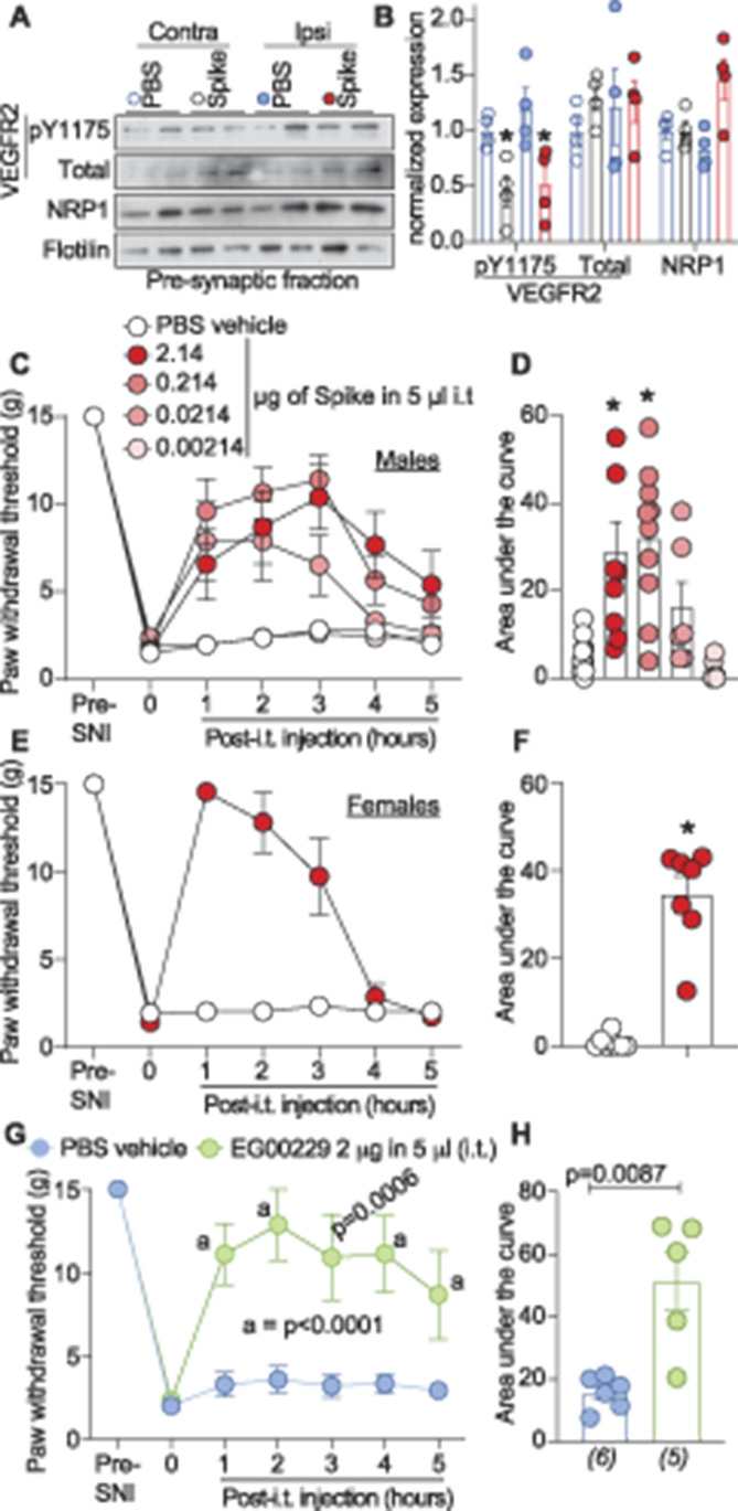
Figure 6.

Severe acute respiratory syndrome coronavirus 2's spike protein and neuropilin-1 receptor (NRP-1) antagonism reverses SNI-induced mechanical allodynia. Spared nerve injury (SNI) elicited mechanical allodynia 10 days after surgery. (A) Representative immunoblots of NRP-1, total and pY1175 VEGF-R2 levels at presynaptic sites in rat spinal dorsal horn after SNI. Tissues were collected 3 hours (ie, at peak antiallodynia) after intrathecal injection of the receptor binding domain of the spike protein (2.14 μg/5 μL). (B) Bar graph with scatter plots showing the quantification of n = 4 samples as in (A) (*P < 0.05, Kruskal–Wallis test). Paw withdrawal thresholds for SNI rats (male) administered saline (vehicle) or 4 doses of the receptor binding domain of the spike protein (0.00214-2.14 μg/5 μL) intrathecally (i.t.); n = 6 to 12) (C) 2.14 μg/5 μL dose of spike in female rats, or the NRP-1 inhibitor EG00229 in male rats (2 μg/5 μL; n = 6) (E). (D, F, H) Summary of data shown in panels C, E, and G plotted as area under the curve (AUC) for 0 to 5 hours. P values, vs control, are indicated. Data are shown as mean ± SEM and were analyzed by nonparametric two-way analysis of variance where time was the within-subject factor and treatment was the between-subject factor (post hoc: Sidak). AUCs were compared by Mann–Whitney test. The experiments were analyzed by an investigator blinded to the treatment. P values of comparisons between treatments are as indicated; for full statistical analyses, see Table S1, http://links.lww.com/PAIN/B190.
