. 2020 Dec 15;9:e60827. doi: 10.7554/eLife.60827

Figure 3. ANT1 inhibitors restore resistance to proton leak of mitochondria in old rat cardiomyocytes.

ANT1 inhibitors 10 μM Bongkrekic Acid (BKA) and 20 μM carboxyatractyloside (CAT), but not 50 μM Genipin (UCP2 inhibitor) or 1 μM oligomycin A (OA, ATPase inhibitor), protected the mitochondrial matrix from decreased pH after exposure to external pH 5.3 (N = 4–19 rats in each group) (A) and reduced the rate of 488/405 decline after exposure to pH 6.9 (N = 4–10 rats in each group) (B). BKA, CAT, Genipin, or OA were added immediately after the mitochondria permeabilization. One-way ANOVA with Fisher’s LSD test was applied. *p<0.05, **p<0.01 vs Young, #p<0.05, ##p<0.01 vs Old.

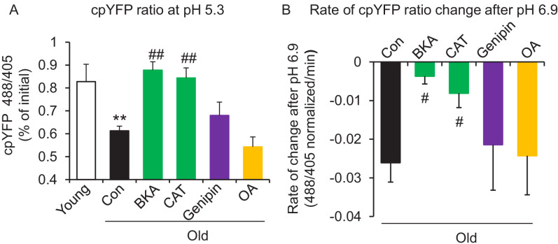
Figure 3.

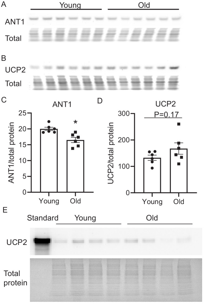
Figure 3—figure supplement 1. Aging effect on ANT1 and UCP2 cardiac protein abundance.

Figure 3—figure supplement 1.

Aging slightly, but significantly decreases ANT1 levels in the mouse heart. Western blot of ANT1 (A, C) and UCP2 (B, D). N = 6 mice in each group. Student’s t-test was applied to determine the statistical significance. *p<0.05 vs young. The total protein loading control was stained with MemCode Reversible Protein Stain Kit for Polyvinylidene difluoride Membranes (Pierce, Rockford, IL). The identity of the band as UCP2 in B was confirmed using a recombinant UCP2 protein standard (Origene, CAT#: TP761109), as shown in E.
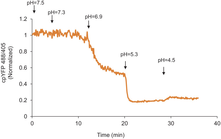
Figure 3—figure supplement 2. Mitochondrial proton entry in buffer without phosphate.

Figure 3—figure supplement 2.

Mitochondrial pH stress on permeabilized rat cardiomyocytes in a buffer that did not contain phosphate.