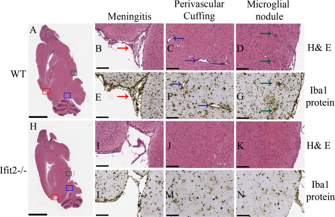
Fig 3. Ifit2 deficiency results in decreased neuroinflammation upon RSA59 infection.
5 μm thin paraffin embedded serial sections of RSA59 infected WT and Ifit2-/- brain tissues processed for H & E (Panels A-D and H-K) and Iba1 staining (Panels E-G and L-N) at 5 days p.i. as indicated. Representative scanned images of whole brain are shown for WT (A) and Ifit2-/- (H) mice with selected meningeal infiltration indicated by Red box, perivascular cuffing by blue box and microglial nodule formation by green box. Selected boxed areas are enlarged for WT and Ifit2-/- brains in Panels B, C, D and Panels I, J, K, respectively. Representative Iba1 staining in meninges, perivascular cuffs and microglial nodules is shown for WT and IFIT2-/- brains in Panels E, F,G and L,M,N, respectively. Ifit2-/- mice showed reduced overall inflammation, reduced or no perivascular cuffing and only scattered Iba-1+ cells without apparent nodule formation. Red arrow depicts menegitis, Dark blue arrow depicts encephalitis (perivascular cuffing), green arrow shows encephalitis (microglial nodules). The experiment was repeated three times and total n = 8. The scale bar for Panel A and H is 2 mm and for Panel B-G and Panel I-N is 75 μm.

