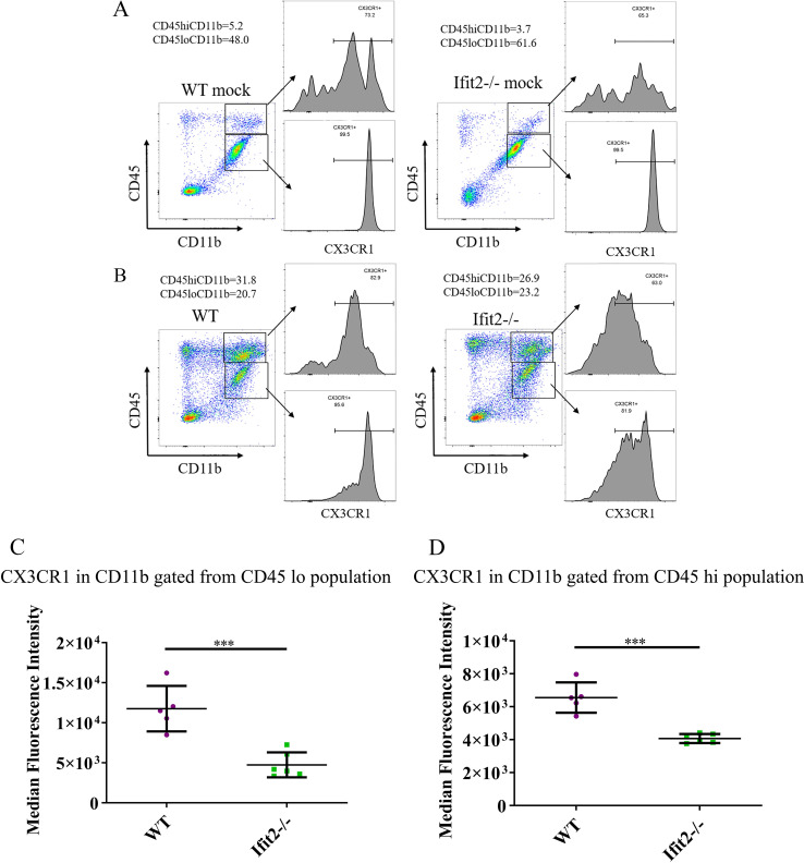
Fig 10. Ifit2 deficiency decreases CX3CR1 expression in microglia following infection.
Brain derived cells from WT and Ifit2-/- mice either mock infected (Panel A) or infected with RSA59 (Panel B) were stained for CD45, CD11b and CX3CR1 expression at 5 days p.i. Dot plots show gating and percentages of CD45hi CD11b+ and CD45lo CD11b+ populations as indicated. Histograms show CX3CR1 expression on the respective gated myeloid cell populations as indicated by arrows. Panels C and D show Median Fluorescent Intensity of CX3CR1 expression by CD45lo CD11b+ microglia and CD45hi CD11b+ infiltrates of WT (Purple) and Ifit2-/- (Green) mice, respectively.The experiment is repeated 2–3 times with n = 5–6. Asterix (*) represents differences that are statistically significant by Student’s unpaired t-test analysis. (***P<0.001).

