
Figure 3. Formation of hASCs spheroids.
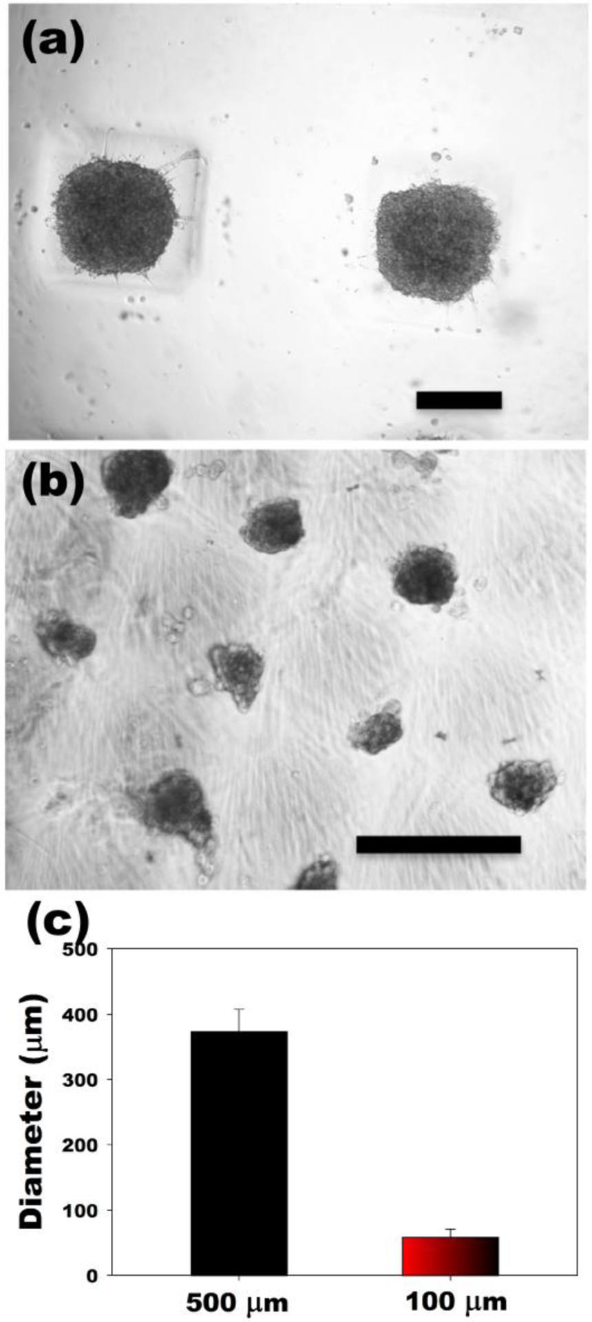
Optical photomicrographs of hASC spheroids in the dual-crosslinked OMA/PEG hydrogel microwells formed with (a) 500 μm and (b) 100 μm grid patterns. (c) Diameter of hASC multicellular spheroids formed in hydrogel microwells. The scale bars indicate 250 μm.