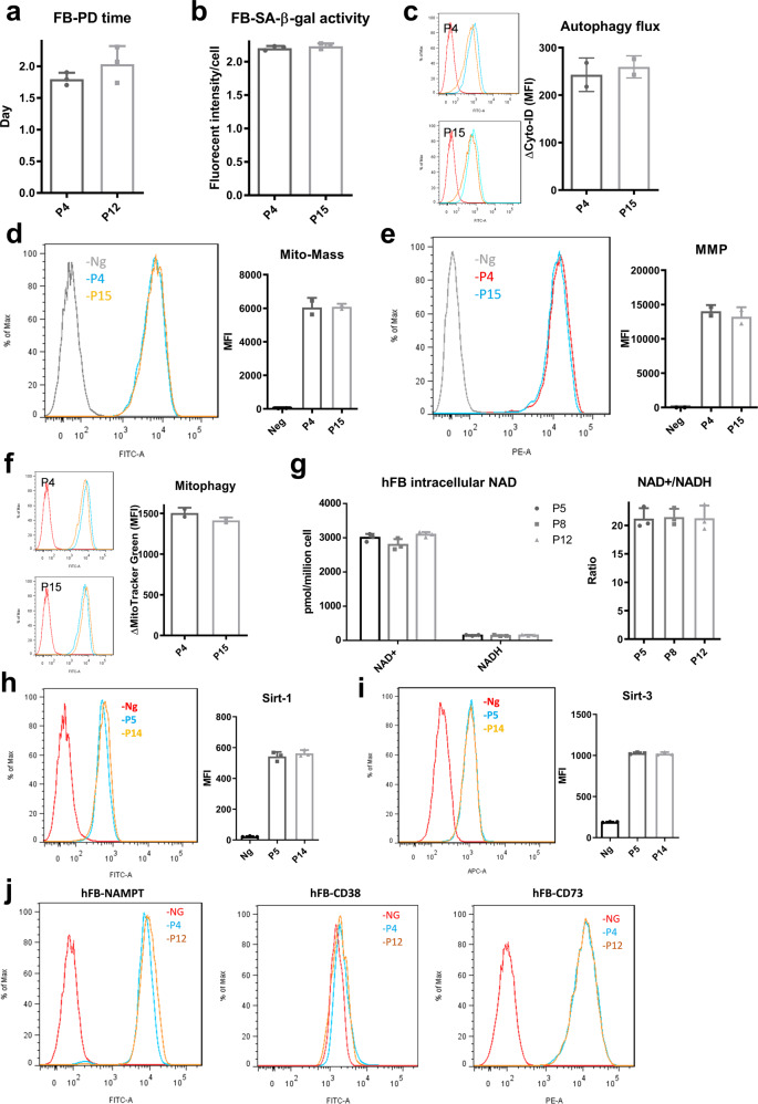
Fig. 7. Human dermal fibroblasts (hFBs) under replicative expansion exhibit limited cellular senescence, mitochondrial dysfunction, and NAD+ decline.
a Population doubling time of hFBs at early and late passage. b SA-β-gal activity for culture-expanded hFBs. c No significant difference for autophagic flux in P4 and P15 hFBs. Red line: negative control. Orange line and blue line represents basal autophagy and with autophagy inhibitor Bafliomycin-A (Baf-A), respectively. d Mitochondrial mass and e mitochondrial transmembrane potential (MMP) showed no difference for P4 and P15 hFBs. f Mitophagy also showed no significant difference in hFBs during culture expansion. Red line: negative control. Orange line and blue line represents untreated and with mitochondrial uncoupler (FCCP) treatment, respectively. g Intracellular NAD+ and NADH levels, as well as NAD+/NADH ratios are relatively stable during culture expansion of hFBs. h Sirt-1 and i Sirt-3 protein expression determined by flow cytometry. j The expressions of NAD+ metabolic enzymes NAMPT, CD38, and CD73 were all comparable for hFBs at different passages as determined by flow cytometry. Biological replicates (n): n = 3.

