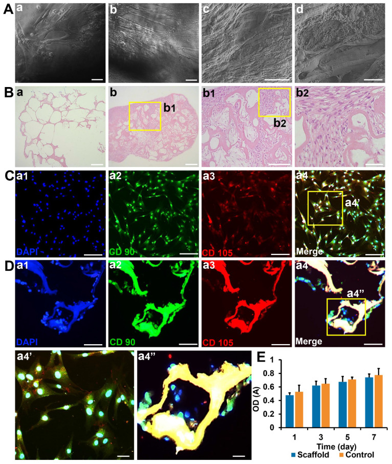
Figure 3.
Biocompatibility and toxicity test of collagen SF scaffold (A) Observation of hUCMSCs cultured on the surface and inside of collagen/SF scaffolds under inverted phase contrast microscopy and SEM. Scale bars: 100 µm (a-c), 20 µm (d). (B) HE staining of blank scaffolds and hUCMSCs co-cultured with collagen/SF scaffolds. Scale bars: 2.5 mm (a-b), 1 mm (b1), 200 µm (b2). (C) Identification of hUCMSCs with CD90 and CD105. Scale bars: 200 µm (a1-a4), 50 µm (a4'). (D) Immunofluorescence staining of surface markers of hUCMSCs when co-cultured with collagen/SF scaffolds. Scale bars: 200 µm (a1-a4), 50 µm (a4''). (E) CCK-8 test of hUCMSCs when co-cultured with collagen/SF scaffolds.

