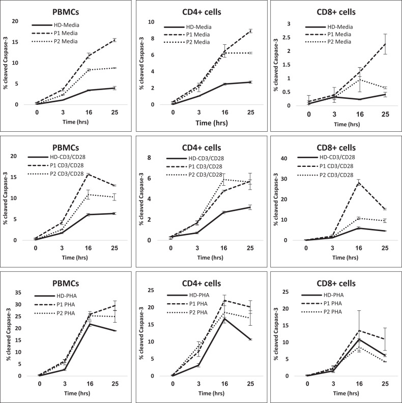
Fig. 2. Stimulation-induced apoptosis.
PBMC apoptosis in response to anti-CD3/CD28 and PHA stimulation. PBMCs from healthydonor (HD, continuous line), P1 (dashed line), and P2 (dotted line) were analyzed for intracellular activated Caspase-3 expression following exposure to anti-CD3/CD28 or PHA for 0, 3, 16, and 25 h. PBMCs (left panels) gated on CD4+ (middle panels) and CD8+ (right panels) were analyzed for cleaved Caspase-3 expression. Significant values were indicated as follows: *p < 0.05; **p < 0.01; ***p < 0.005.

