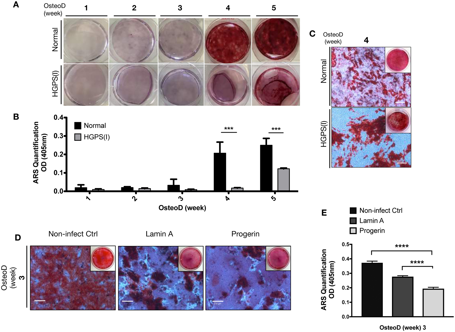
Figure 1: Osteoprogenitor cells with HGPS mutation undergo defective osteogenic differentiation.

(A) Normal and HGPS iPSC-osteoprogenitors were induced for Osteogenic differentiation (OsteoD) for up to five weeks, and the calcium depositions in the cells were evaluated by Alizarin Red S (ARS) staining assay. Representative images are shown. (B) Quantification of ARS staining in normal and HGPS osteoprogenitors (n=3). (C) Representative whole plate views and magnified bright-field microscopy images of normal and HGPS osteoprogenitors that were differentiated for four weeks, and stained with ARS. Images are taken from biological replicate of (A). Scale bars, 200μm. (D) Representative whole plate views and magnified bright-field images of ARS stained hBM-MSCs with no lentiviruses (Control), or lentivirus-mediated overexpression of wild-type lamin A or progerin. Scale bars, 200μm. (E) Quantification of colorimetric detection of ARS stains (n=3) from samples in (D). Data are represented as mean ± SD. ***p < 0.001, ****p < 0.0001.
