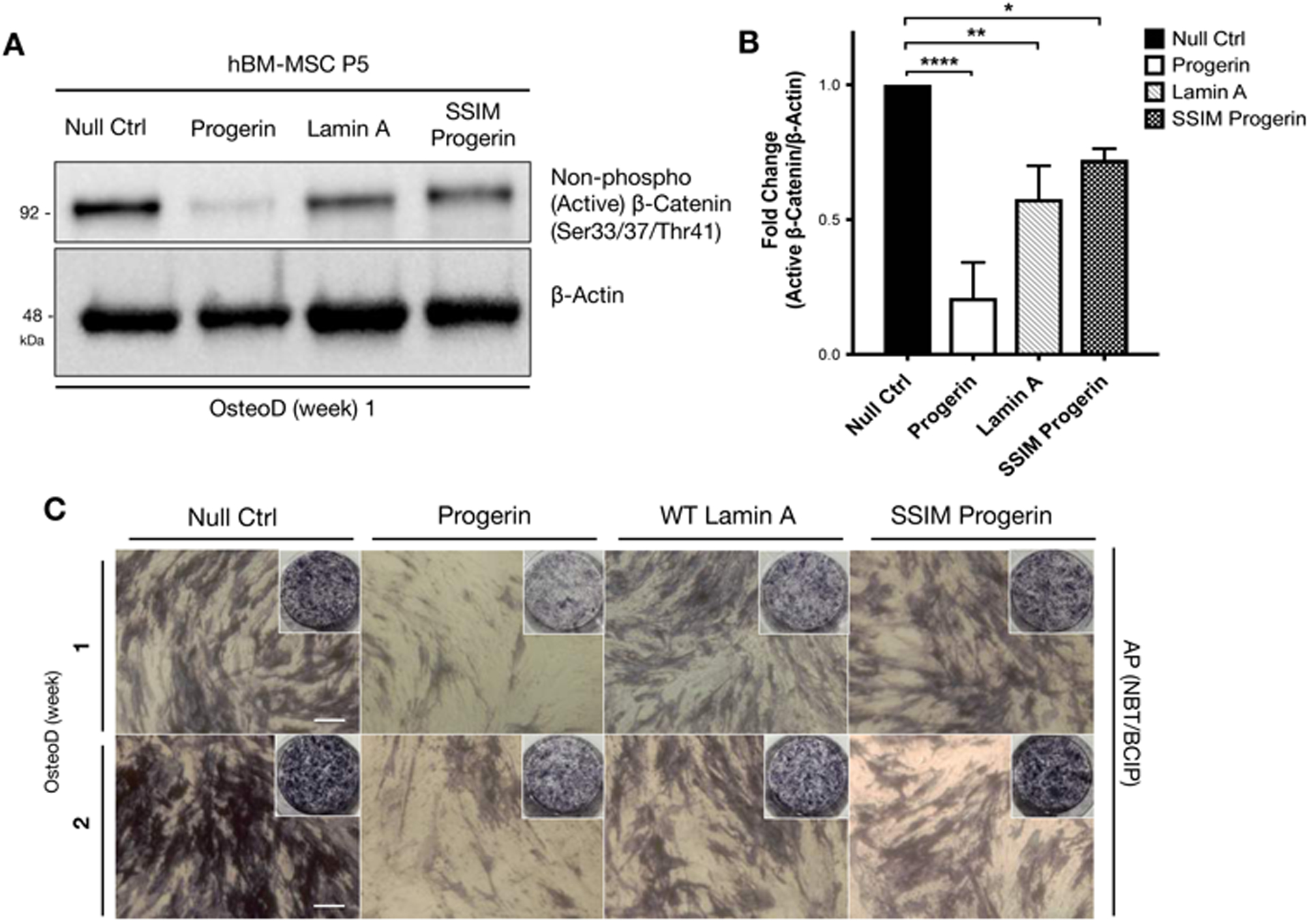
Figure 3: Wild-type hMSCs overexpressing a non-farnesylable form of progerin attain stabilized active β-catenin activity during the osteogenic differentiation.

(A) Representative Western blot detecting non-phosphorylated (active) β-catenin in hBM-MSCs induced with lentiviruses expressing GFP, GFP-progerin, GFP-lamin A, or GFP-SSIM-progerin. β-actin protein was used as a loading control. The samples were collected at one week in osteogenic differentiation. (B) Quantification of western blot by analyzing the fold changes of non-phosphorylated (active) β-catenin/β-actin densitometry. Western blots were conducted at least three biological replicates (n>3) on separate gels. (C) Whole plate views and bright-field images of alkaline phosphatase (AP) staining in hBM-MSCs overexpressing GFP-null, GFP-progerin, GFP-lamin A and GFP-SSIM (non-farnesylable) progerin, then induced for osteogenic differentiation for one or two weeks.
