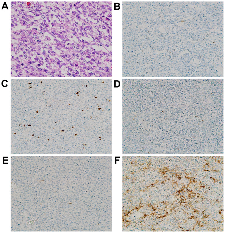
Figure 3.
Photomicrographs of the small intestinal tumor. (A) A patternless proliferation of atypical cells characterized by scant pale cytoplasm with microvesicular degeneration (H&E stain; magnification, x200). The tumor was immunohistochemically partially positive for (B) S-100 (IHC stain; magnification, x100) and (C) SOX10 (IHC stain; magnification, x100), but negative for (D) HMB45 (IHC stain; magnification, x100) and (E) Melan A (IHC stain; magnification, x100). Additionally, the tumor was positive for (F) CD56 (IHC stain; magnification, x100). IHC, immunohistochemical; SOX10, SRY-related HMG-box 10; HMB45, human melanin black-45.

