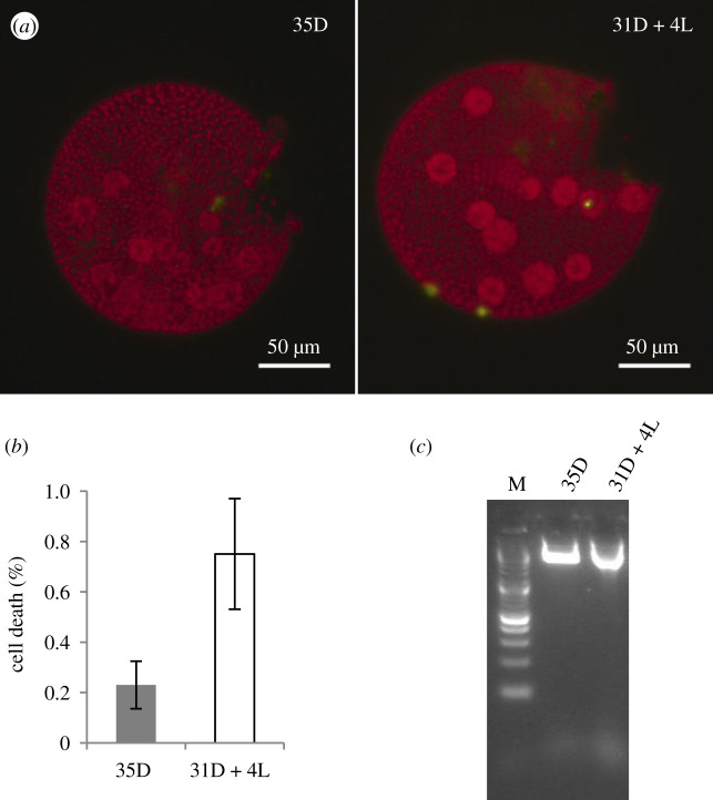
Figure 7.
The effect of light following an extended dark period on the viability of juvenile EVE somatic cells. EVE cultures were subjected to 31D + 4L and 35D as in figure 6a. (a) Representative fluorescent micrographs of EVE juveniles subjected to 35 h of dark (35D) or 31 h of dark followed by 4 h of light (31D + 4L); dead cells appear green (SYTOX Green) and live cells are red (due to chlorophyll autofluorescence). (b) Percentage of dead somatic cells (per individual) from 35D and 31D + 4L cultures (n = 3; 3 technical replicates with greater than or equal to 20 individuals each; bars indicate s.e.); (c) total DNA extracted from 35D and 31D + 4L cultures not showing the DNA laddering effect characteristic of PCD. M, DNA marker.

