
Figure 1: Mural cell markers are enriched in human and murine brain microvessel extracts.
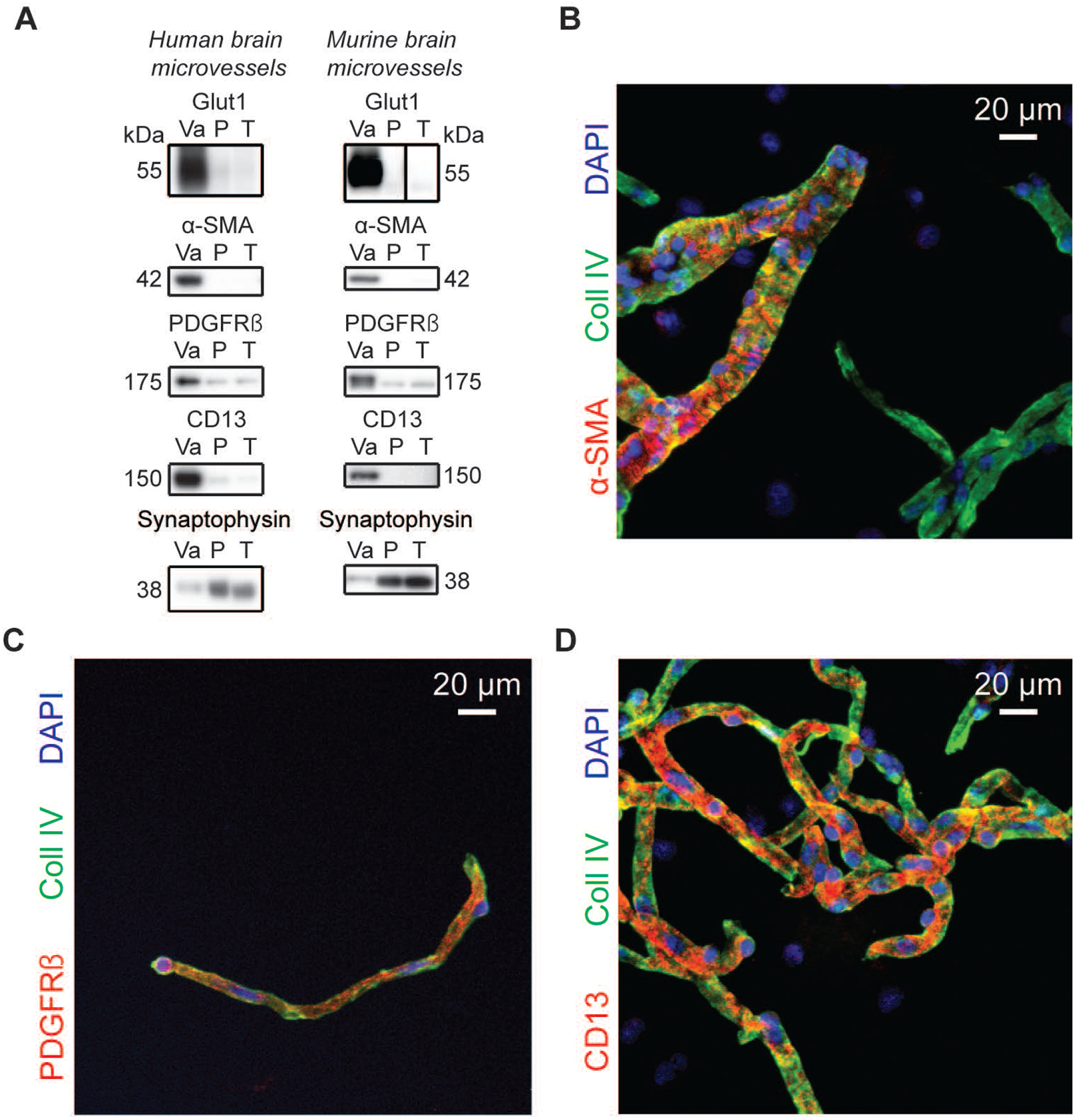
A) Western immunoblotting analyses on human and murine brain microvessel extracts show that the endothelial marker Glut1 as well as known mural cell markers are concentrated in the vascular fraction whereas synaptophysin, a neuronal marker, is enriched in the microvessel-depleted parenchymal fraction. Representative photo examples were taken from the same immunoblot experiment, and black vertical line was inserted to indicate nonconsecutive bands. The same amount (8 μg) of proteins per sample was loaded. B-D) Subsequent immunofluorescence assays on human brain microvessels showed that antibodies raised against α-SMA labeled larger vessels while capillary-like smaller vessels were stained by PDGFRβ and CD13 antibodies. Scale bar: 20 μm. Abbreviations: α-SMA, smooth muscle alpha actin; CD13, aminopeptidase N; Coll IV, type IV collagen; P, microvessel-depleted parenchymal fraction; PDGFRβ, platelet-derived growth factor receptor β; T, total homogenate; Va, vascular fraction, enriched in microvessels.