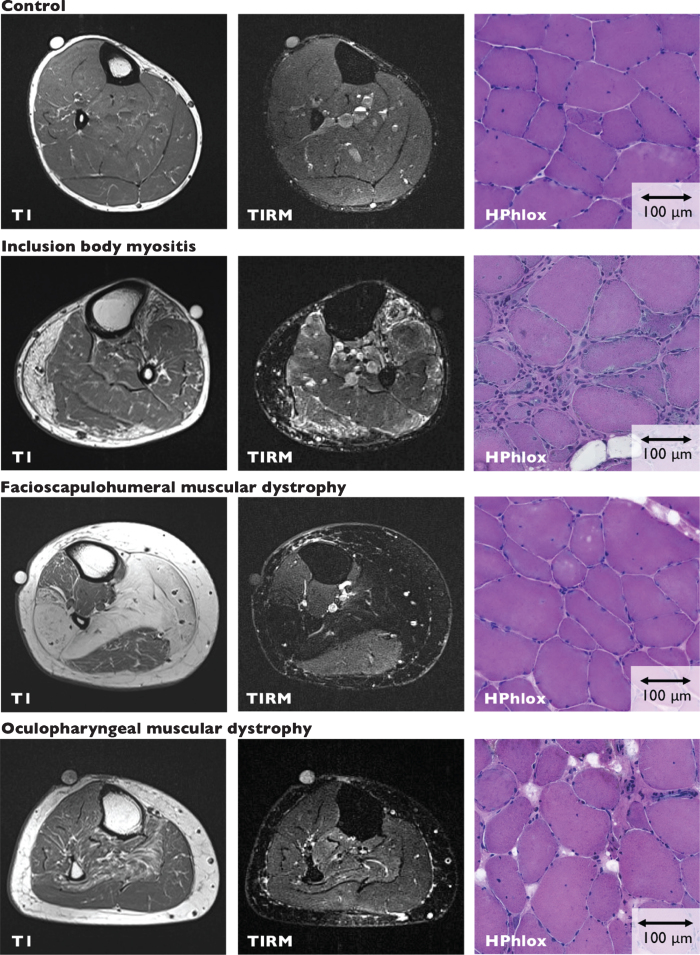
Fig. 1.
MRI imaging and muscle biopsy sections. Representative T1 and TIRM images and corresponding muscle biopsy sections (HPhlox staining) from the tibialis anterior of control, IBM, FSHD and OPMD participants. Control Participant C3, MRI shows 3% fatty infiltration and negative TIRM. The muscle biopsy has a histopathological severity sum score of 3 and shows mildly increased variability in fiber size, a mild increase in internal nuclei, no necrosis and/or regeneration, and mild fibrosis. Inflammation was scored as none. IBM Participant I2, MRI shows fatty infiltration and TIRM-hyperintense changes. Quantitative assessment of fatty infiltration is not reliable in TIRM hyperintense muscles. The muscle biopsy has a histopathological severity sum score of 11 and shows severely increased variability in fiber size, a moderate increase in internal nuclei, severe necrosis and/or regeneration, and severe fibrosis. Inflammation was scored as severe. FSHD Participant F2, MRI shows 20% fatty infiltration and negative TIRM. The muscle biopsy has a histopathological severity sum score of 7 and shows moderately increased variability in fiber size, a moderate increase in internal nuclei, mild necrosis and/or regeneration, and moderate fibrosis. Inflammation was scored as none. OPMD Participant O14, MRI shows 3% fatty infiltration and negative TIRM. The muscle biopsy has a histopathological severity sum score of 5 and shows mild increased variability in fiber size, a moderate increase in internal nuclei, mild necrosis and/or regeneration, and mild fibrosis. Inflammation was scored as none.

