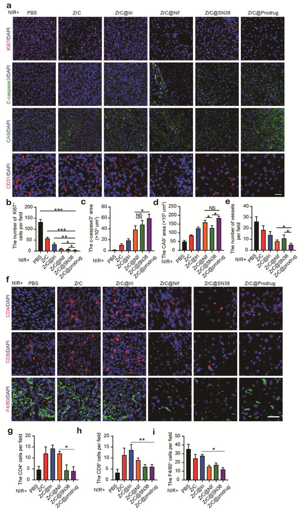
Figure 5.

Synergistic inhibition of tumor growth with dual targeting malignant cells and tumor stroma. Tumors tissues in colorectal liver metastasis (CRLM) model, were isolated 14 days after various treatments, and a) representative immunofluorescence staining analysis of Ki67, C‐caspase3, CA9 and CD31 in tissues were obtained for the changes in the tumor environment (TME). Statistical analysis of a functional parameter type, such as b) cancer cell proliferation by Ki67, c) cellular apoptosis by C‐caspase3, and d) hypoxia signal by CA9+, and e) regressive vasculature by CD31. f) Representative immunofluorescence staining analysis of CD4, CD8, and F4/80 in CRLM tumor tissues. g) Statistical analysis of CD4, CD8, and F4/80‐positive cells in each field. n > 5 fields from four tumors. Bar graphs show the mean ± SD. *p < 0.05, **p < 0.01, and ***p < 0.001. ns: no significance. Scale bar, 50 µm.
