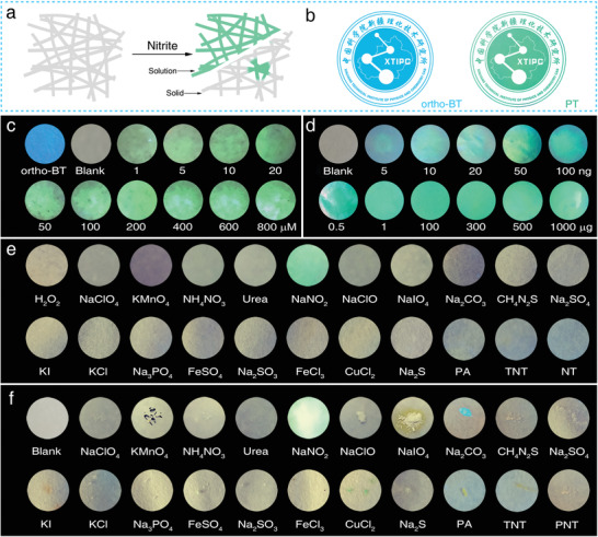
Figure 3.

a) Schematic of the test strips used to detect a nitrite solution and solid particles. b) Optical images of the special logo stamped by ortho‐BT and PT. Optical images of the test strips used to detect c) a nitrite solution (0–800 × 10−6 m) and d) nitrite solid (0–1 mg). Optical images of the anti‐interference characterization of the test strips to >20 types of substances in the e) solution and f) solid phase, respectively. All images were obtained under irradiation with a 365 nm UV lamp and all the image diameters in (c)–(f) were maintained at 10 mm. (Excitation source: 365 nm LED; Exposure time: 1/40 s).
