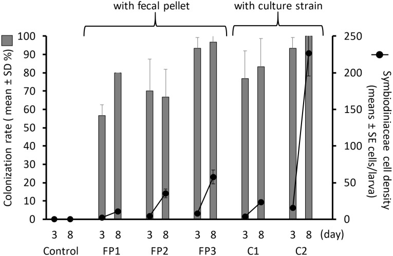
Fig 4. Proportions of Acropora tenuis larvae colonized with symbiont cells (bars) and colonizing cell densities per larva (lines with dots).
FP1, FP2 and FP3 represent the experimental groups larvae provided fecal pellets from different individuals of Tridacna crocea. C1 and C2 refer larvae supplied with a mixture of AJIS2-C2 (Symbiodinium microadriaticum) and CCMP2556 (Durusdinium trenchii) and a mixture of TsIs-H4 (S. tridacnidorum) and TsIs-G10 (Durusdinium sp.), respectively. The control group was not provided any symbiont source.

