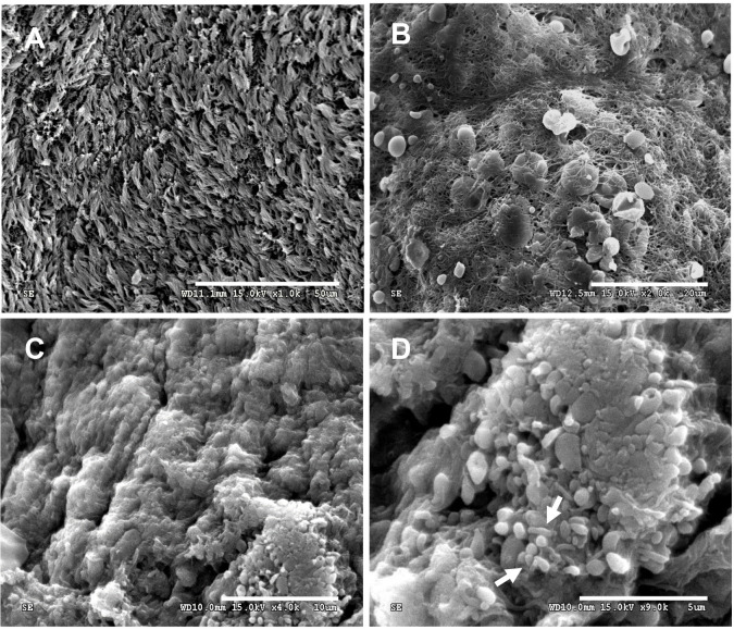
Fig 1. Scanning electron micrographs of mucosal tissues from CRS patients.
(A) Maxillary sinus mucosa without evidence of biofilm but positive for bacteria when cultured; bar = 50 μm. (B) Wire-like structures seen on the sinus mucosal epithelium; bar = 20 μm. (C) Large biofilm aggregates; bar = 10 μm and (D) biofilm with coccus- and bacillus-shaped elements (white arrows); bar = 5 μm.

