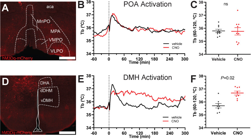
Fig 7. Chemogenetic activation of DMHAdora1, but not POAAdora1 neurons increases Tb.
A-C tests activation of the POA. (A) Example of hM3Dq-mCherry expression in Adora1-Cre mouse injected with AAV8-hSyn-DIO-hM3Dq-mCherry in the POA. (B) Tb response to CNO (1 mg/kg, i.p.) vs. vehicle in Adora1-Cre mice with hM3Dq-mCherry in POA (n = 11). (C) Mean Tb at 60 to 120 minutes after dosing with CNO (1 mg/kg, i.p.) vs. vehicle. D-F tests activation of the DMH. (D) Example of hM3Dq-mCherry expression in Adora1-Cre mouse injected with AAV8-hSyn-DIO-hM3Dq-mCherry in the DMH. (E) Tb response to CNO (1 mg/kg, i.p.) vs. vehicle in Adora1-Cre mice with hM3Dq-mCherry in DMH (n = 8). (F) Mean Tb at 60 to 120 minutes after dosing with CNO (1 mg/kg, i.p.) vs. vehicle (n = 8). Data are mean ± SEM (SEM is omitted for visual clarity in B and E). P values were calculated by paired t-test comparing drug vs vehicle within mouse. Scale bar is 500 μm. aca, anterior commissure; dDMH, dorsomedial hypothalamus, dorsal part; DHA, dorsal hypothalamic area; MnPO, median preoptic area; MPA, medial preoptic area; vDMH, dorsomedial hypothalamus, ventral part; VMPO, ventromedial preoptic area; VLPO, ventrolateral preoptic area.

