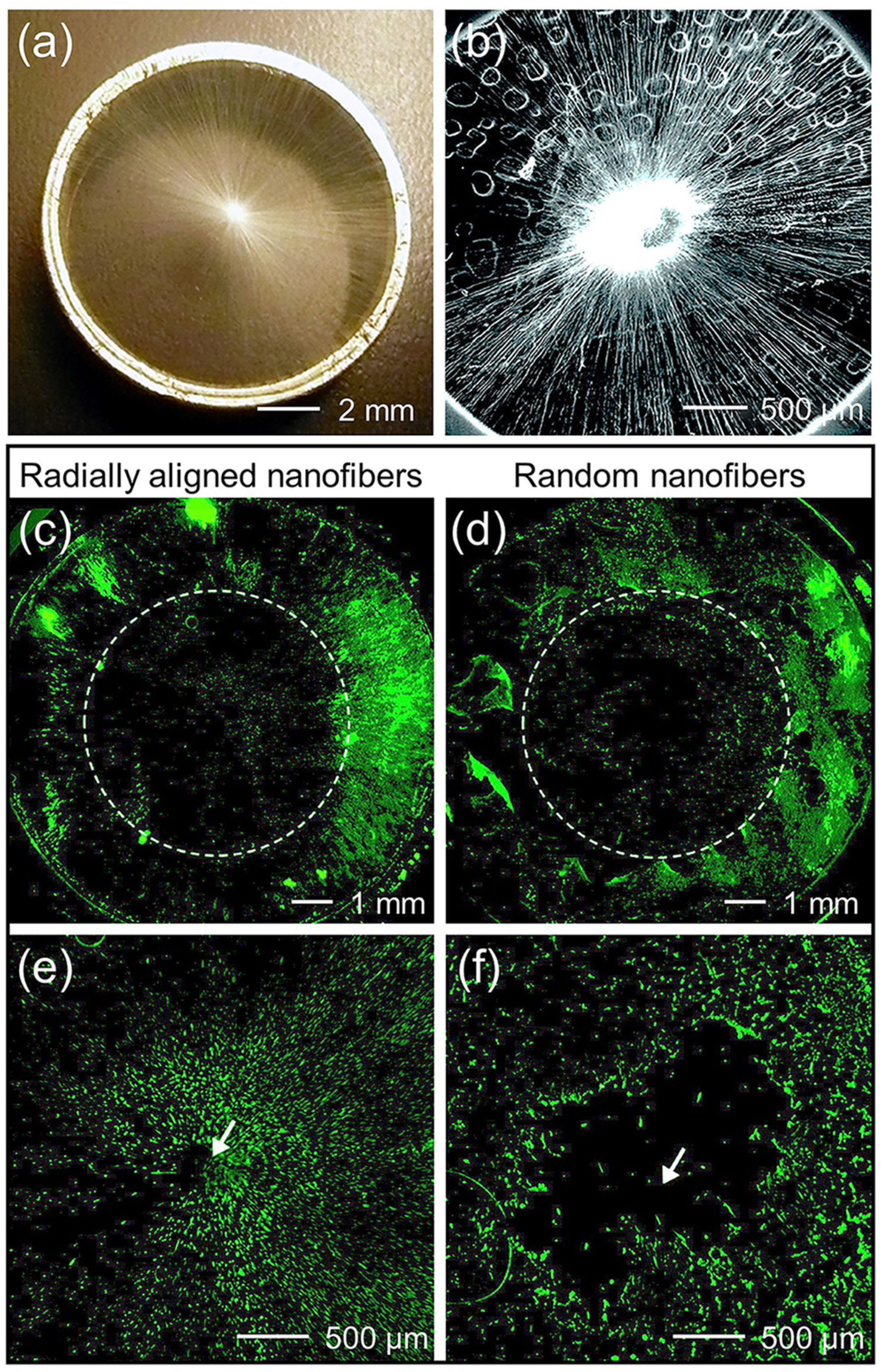
FIG. 6.

Migratory behavior of cells on radially aligned nanofibers. (a) Photograph and (b) SEM image showing the radial alignment of PCL nanofibers in a scaffold that was directly deposited on the ring collector. Fluorescence micrographs showing the migration of dura fibroblasts on scaffolds made of [(c) and (e)] radially aligned and [(d) and (f)] random PCL nanofibers from the periphery of the scaffold toward the center after four days of incubation. Enlarged views of the center portions in (c) and (d) are shown in (e) and (f), respectively. The arrow marks the center of the scaffold.49 Reprinted with permission from Xie et al., ACS Nano 4, 5027 (2010). Copyright 2010 American Chemistry Society.
