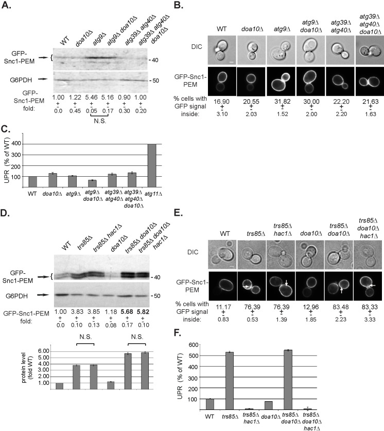
Fig 6. doa10Δ does not increase the constitutive ER-phagy defect of atg9Δ mutant cells, and its effect on trs85Δ phenotype is not dependent on UPR.
A-C. Effect of doa10Δ alone and in combination with atg9Δ and atg39Δ atg40Δ. WT and indicated mutant cells overexpressing GFP-Snc1-PEM were grown in normal growth medium (SD+N). The level of GFP-Snc1-PEM in cell lysates was determined by immuno-blot analysis using anti-GFP antibodies (A); intracellular accumulation of GFP-Snc1-PEM was determined by live-cell fluorescence microscopy (B); and UPR induction was analyzed (C). Results are presented as in Fig 2. doa10Δ has no effect on GFP-Snc1-PEM accumulation in combination with atg9Δ, which exhibits a constitutive ER-phagy phenotype, or atg39Δ atg40Δ double deletion, which does not exhibit a constitutive ER-phagy phenotype. D-F. Abrogation of the UPR response by hac1Δ does not affect the constitutive ER-phagy phenotype of trs85Δ, or its enhancement in trs85Δ doa10Δ mutant cells. WT and indicated mutant cells overexpressing GFP-Snc1-PEM were grown in normal growth medium (SD+N). The level of GFP-Snc1-PEM in cell lysates was determined by immuno-blot analysis using anti-GFP antibodies (D); intracellular accumulation of GFP-Snc1-PEM was determined by live-cell fluorescence microscopy (E); and UPR induction were analyzed as explained for Fig 2C (F). The 50% increase in the GFP-Snc1-PEM accumulation phenotype in trs85Δ doa10Δ double mutant cells is affected by hac1Δ (panel D). Results are presented as in Fig 2 and represent 3 independent experiments.

