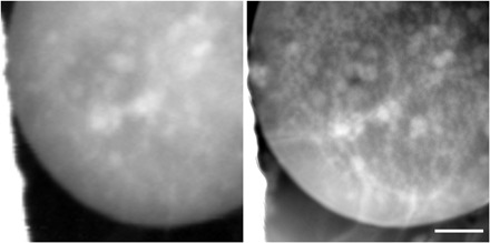
Fig. 4. Cryogenic microscopy of a frozen hydrated yeast cell.

Conventional (left) and ptychographic (right) imaging of a frozen hydrated yeast cell using 520-eV x-rays. The ptychographic image shows scattering contrast and demonstrates improved contrast and resolution compared to conventional imaging. Scale bar, 1 μm; reconstructed pixel size, 5 nm.
