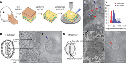
Fig. 1. The bill skin of a tactile-specialist duck has Pacinian and Meissner corpuscles.

(A) Schematic illustration of the preparation of duck bill skin for electrophysiological and optical analysis of mechanosensory corpuscles. (B) A bright-field microscopic image of a mixed population of Pacinian corpuscles (blue arrowheads) and Meissner corpuscles (red arrowheads) in a patch of duck skin from the dorsal surface of the upper bill. (C) Size distribution of visible Meissner and Pacinian corpuscles in duck bill skin (50 Meissner and 140 Pacinian corpuscles in total). (D to I) Illustrations (D and G), electron microscopy images (E and H), and close-up bright-field microscopy images (F and I) of mechanosensory corpuscles. Pacinian corpuscles are composed of outer core lamellar cells surrounding an inner bulb of inner core cells and a neuronal mechanoreceptor. In Meissner corpuscles, the mechanoreceptor is sandwiched between two or more lamellar cells.
