
Fig. 2. Lamellar cells of Pacinian and Meissner corpuscles are mechanosensitive.
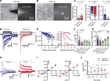
(A and B) Representative bright-field (left) and fluorescent images (right) of lamellar cells from Pacinian and Meissner corpuscles filled with Lucifer yellow via the recording electrode. A glass probe is positioned nearby to deliver mechanical stimulation. (C) Electrophysiological characteristics of lamellar cells. Significance was calculated using unpaired two-tailed t test. Open circles denote individual cells. (D) Representative MA currents elicited from lamellar cells by mechanical indentation using a glass probe. (E) Quantification of peak MA current amplitude in Pacinian (left, n = 19 cells) and Meissner (right, n = 7 cells) lamellar cells in response to indentation with a glass probe. Lines connect measurements from individual cells. (F) Quantification of MA current rise time (τrise) recorded in lamellar cells and in duck trigeminal mechanoreceptors with fast, intermediate, and slow MA current. The difference between means is significant; F4,61 = 3.49, P = 0.013, one-way analysis of variance (ANOVA) with Tukey’s post hoc multiple comparisons test. Open circles denote individual cells. (G) Quantification of MA current inactivation rate (τdecay) recorded in lamellar cell and duck trigeminal mechanoreceptors. τdecay values greater than 1000 ms are plotted as 1000. Bars represent median. Open circles denote individual cells. The difference between medians is significant; P < 0.0001, Kruskal-Wallis test. ***P < 0.0001, Dunn’s post hoc multiple comparisons test. (H) Representative MA currents elicited from lamellar cells in response to indentation at different voltages. (I) Voltage dependence of peak MA current from eight Pacinian and five Meissner lamellar cells, fitted to the linear equation. (J) Quantification of MA current τdecay from seven Pacinian and seven Meissner lamellar cells, fitted to the linear equation. r, Pearson’s correlation coefficient; P denotes the probability of the line slope being equal to 0. Data are presented as means ± SEM from at least three independent skin preparations.