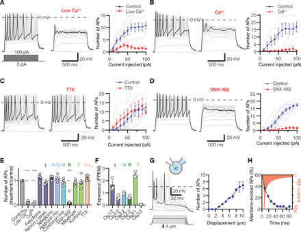
Fig. 4. Lamellar cells from Meissner corpuscles are excitable mechanosensors.

(A to D) Exemplar action potentials (APs; left and middle) and spike quantification (right) obtained by current injection into Meissner lamellar cells in the presence of low (20 μM) extracellular Ca2+ [(A), n = 6 cells], pan-Cav blocker Cd2+ [(B), n = 4 cells], Nav blocker tetrodotoxin (TTX; (C), n = 5 cells], R-type Cav2.3 blocker SNX-482 [(D), n = 11 cells]. Thin lines in quantification panels represent individual cells; thick lines connect means ± SEM. (E) Pharmacological profile of Meissner lamellar cell firing upon injection of a 100-pA current. Letters indicate Cav type. Open circles represent individual cells. The effect of treatment is significant. F11,53 = 75.57, P < 0.0001, one-way ANOVA; ***P < 0.0001 versus control, Dunnett’s test. (F) Quantification of Cav alpha subunit expression in duck bill skin (n = 3 animals). FPKM, number of mRNA fragments per kilobase of exon per million fragments mapped. (G) Mechanical stimulation evokes action potential firing in Meissner lamellar cells. Shown are exemplar action potential traces (left) and quantification of mechanically evoked firing (right) from three Meissner lamellar cells (thin lines). Thick line connects means ± SEM. (H) The number of action potentials is maximal when MA current is at its peak. Shown is a cumulative frequency distribution of the number of action potentials (circles) evoked by mechanical stimulation of four Meissner lamellar cells to 8-μm depth, fitted to the single exponential equation (line) and plotted against peak-normalized MA current profile.
