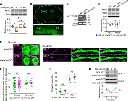
Fig. 7. KIF17 3′UTR is essential for KIF17 synthesis induced by fear memory retrieval.

(A) Time course of KIF17 expression in the mouse hippocampus after contextual fear memory retrieval. HC, home cage mice. In box plots, the central line represents the median, the edges of the box represent the interquartile range, and whiskers represent the minimum to the maximum. P < 0.01, one-way ANOVA; *P < 0.05, **P < 0.01, Bonferroni’s post hoc comparison. n = 5 mouse groups. See also fig. S9. (B) Representative images of a coronal brain section from the mouse bilaterally microinjected AAV-GFP into the hippocampi. Tile scanning with an LSM780 confocal laser scanning microscope was used. The box in the top panel represents the magnified region in the bottom panel. DG, dentate gyrus. Scale bars, 500 μm. See also fig. S10 (A to D). (C) Genomic PCR of the indicated hippocampal lysates. bp, base pair. (D) Comparison of the endogenous KIF17 levels in the hippocampus. P ≥ 0.05, one-way ANOVA. n = 4 mouse groups. WT, wild-type. (E and F) Representative images of immunofluorescence histochemistry demonstrating KIF17 expression in AAV-infected neurons (left, somata; right, dendrites) at the CA3 region of coronal brain sections (E) and quantification of the normalized density of anti-KIF17–positive puncta in somata and dendrites (F). Scale bars, 10 μm. NS, P ≥ 0.05, two-tailed t test. n = 25 to 29 somata and 11 dendrites from three mouse pairs. See also fig. S10 (E to H). (G) Freezing scores of the contextual fear conditioning test. NS, P ≥ 0.05, two-tailed t test. n = 6 mouse pairs. (H) Immunoblotting demonstrating the specific disruption of the synthesis of KIF17 by AAV-Cre–mediated deletion of Kif17 3′UTR. Effects of treatment (P < 0.05) and time (P < 0.01), two-way ANOVA; **P < 0.01, Bonferroni’s post hoc comparison. n = 3 mouse groups.
