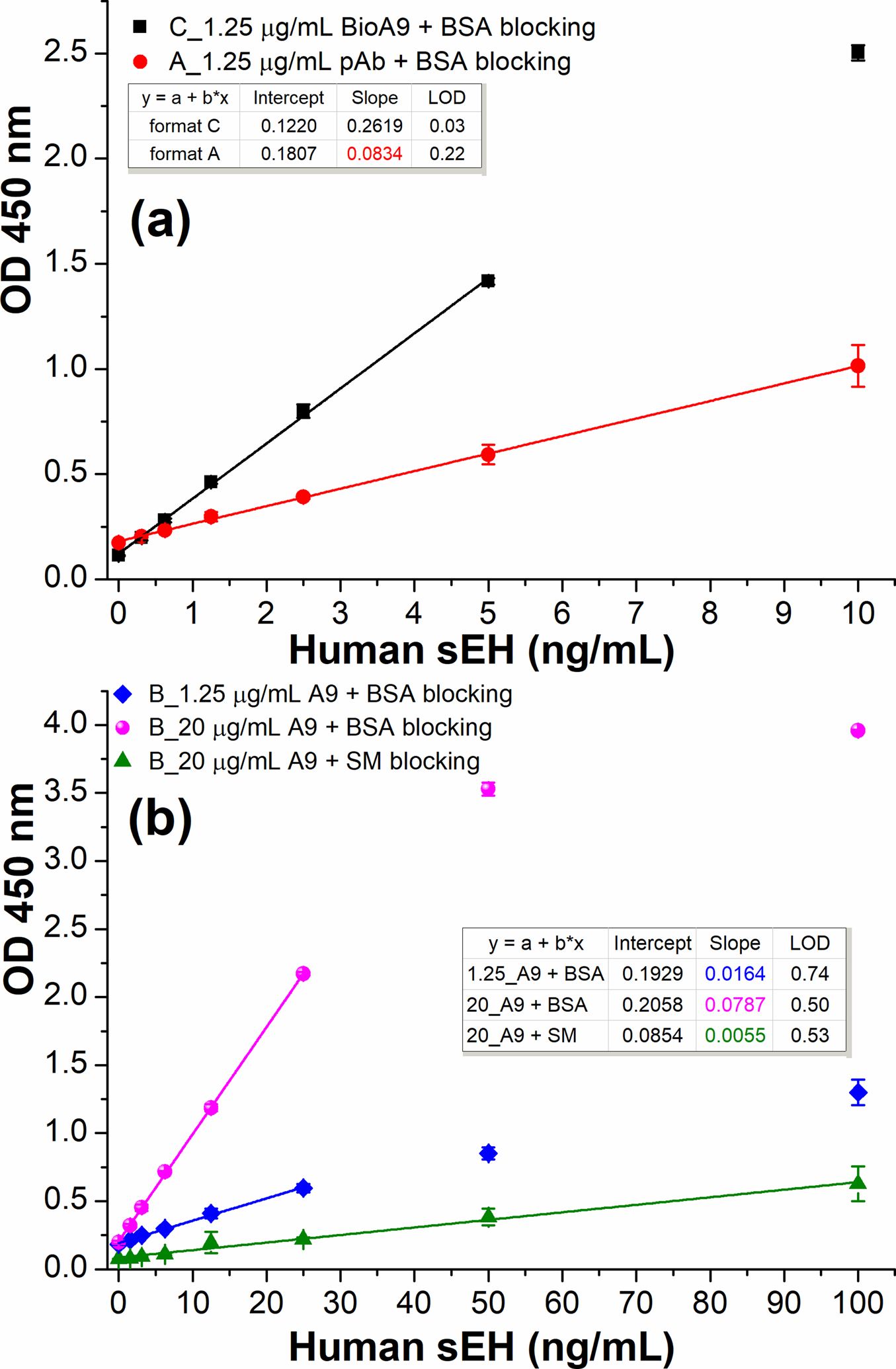
Figure 5.

Comparison of three ELISA formats using HRP-A1 (1:6400 dilution in PBS) as detection antibody. (a) format C (SBdNb ELISA, square): 2.5 μg/mL streptavidin, 2% BSA/PBS blocking, 1.25 μg/mL biotin-A9; format A (circle): 1.25 μg/mL anti-sEH pAb, 2% BSA/PBS blocking. (b) format B with varying nanobody A9 coated in PBS and different blocking agents: diamond, 1.25 μg/mL A9 plus 2% BSA/PBS blocking; sphere, 20 μg/mL A9 plus 2% BSA/PBS blocking; up-triangle, 20 μg/mL A9 plus 3% SM/PBS blocking. All same or similar steps were performed under the same conditions. Error bars indicate standard deviations (n = 3). All R2>0.99 except the up-triangle of Fig. 5b (R2=0.9878).
