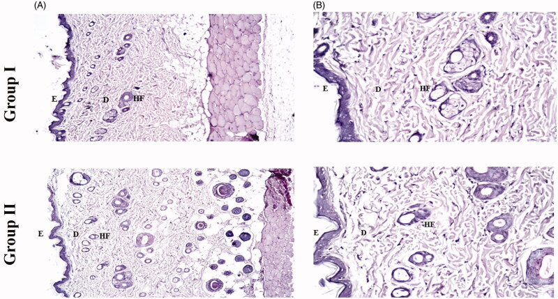
Figure 4.
Photomicrographs showing histopathological sections (hematoxylin and eosin stained) of rat’s skin normal control (group I) and rat’s skin treated with PC6 (group II) with magnification power of 16x to illustrate all skin layers (A) and magnification power of 40x to identify the epidermis and dermis (B). E: epidermis; D: dermis; HF: hair follicles; FTN: fenticonazole nitrate; PC: PEGylated cerosomes.

