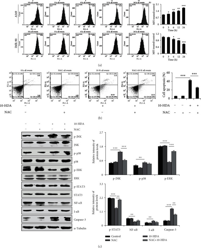
Figure 4.

Effects of ROS generation and apoptosis after treatment of human lung cancer cells with 10-HDA. (a) A549 cells and IMR90 human normal lung fibroblasts were treated with 30 μM 10-HDA after 24 h. ROS levels were examined by flow cytometry. (b) A549 cells were cultured with 30 μM 10-HDA or 0.25 μM (20 μL/mL) NAC for 24 h, and cell apoptosis was detected by flow cytometry analysis. (c) A549 cells were pretreated with 0.25 μM (20 μL/mL) and NAC for 30 min, followed by treatment with 10-HDA for 24 h. The expression levels of MAPK, STAT3, NF-κB, and caspase-3 were detected by western blot analysis and normalized to α-tubulin. The expression levels of proteins were analyzed with ImageJ software. ∗p < 0.05, ∗∗p < 0.01, and ∗∗∗p < 0.001 vs. the control group and the NAC+10-HDA group.
