
Figure 2.
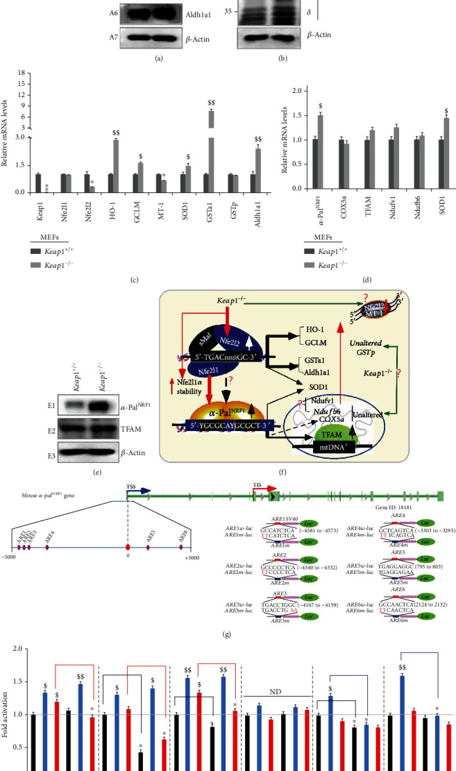
Distinct roles of key redox control genes in the basal expression of mouse α-PalNRF1, TFAM, and relevant genes. (a) Distinct protein levels of keap1 and Nfe2l2, as well as indicated antioxidant enzymes, in between Keap1−/− and Keap1+/+ MEFs were visualized by Western blotting. (b) Altered abundances of distinct Nfe2l1 isoforms between Keap1−/− and Keap1+/+ MEFs were observed. (c) Basal mRNA levels of keap1, Nfe2l1, and Nfe2l2, as well as indicated antioxidant genes, were determined by real-time qPCR of Keap1−/− and Keap1+/+ MEFs. The data are shown as mean ± SEM (n = 3 × 3) with significant decreases (∗p < 0.01, ∗∗p < 0.001) or significant increases ($, p < 0.01; $$, p < 0.001). (d) Basal mRNA levels of α-PalNRF1, COX5a, TFAM, Ndufv1, Ndufb6, and SOD1 were also determined as described above. (e) Altered protein levels of α-PalNRF1 and TFAM were revealed by Western blotting of Keap1−/− and Keap1+/+ MEFs. (f) A model is proposed to present effects of Keap1−/− on Nfe2l1, Nfe2l2, α-PalNRF1, TFAM, and related genes (and their proteins) in MEFs. (g) Within the promoter region of mouse α-PalNRF1 gene, the putative ARE sites (each with the core sequence 5′-TGAC/GnnnGC-3′) were marked (as purple dots, left panel). Six distinct ARE-driven reporters (i.e., ARE1 to ARE6-luc) and their respective mutants were constructed into the pGL3-Promoter vector (right panel). (h) Each pair of indicated ARE-luc and mutants was cotransfected with the internal control pRL-TK, together with each of expression constructs for mouse Nfe2l1, Nfe2l2, or empty pcDNA3.1 into RL34 cells for 8 h, before being allowed for 24 h recovery. Subsequently, distinct ARE-driven luciferase activity was measured. The resultant data are shown as mean ± SEM (n = 3 × 3) with significant increases ($, p < 0.01; $$, p < 0.001) or decreases (∗p < 0.01). ND: no statistical difference.