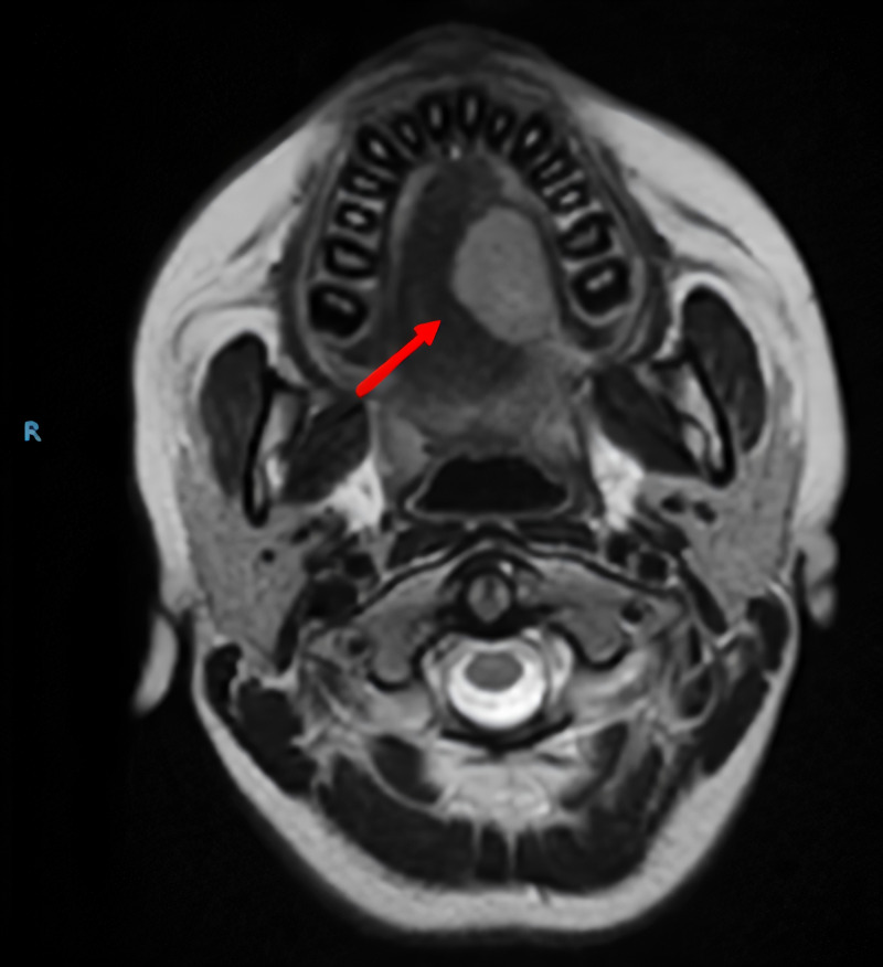
Figure 2. Magnetic resonance imaging of the face and neck.
Involvement of the mucosa and the superior longitudinal, vertical, and transverse muscles of the lateral dorsum of the left hemilanguage was observed by a mass of well-defined and regular contours with increased intensity of signal in the T2-weighted sequences, with intense and homogeneous enhancement with the contrast medium, with measurements of 21 mm x 26 mm x 21 mm.

