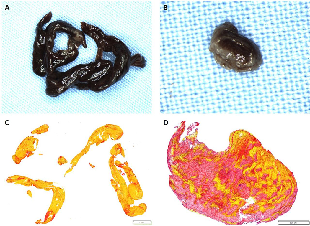
Fig. 3:

Gross photographs and histological staining of removed clots. Gross photographs of a RBC-rich and Fibrin-rich clot (A (1.25x) and B (3.2x), respectively) were taken after formalin-fixation, prior to embedding. C and D are the corresponding MSB-stained slides demonstrating the presence of Red Blood Cells (Yellow), White Blood Cells (Blue), fibrin strands (Red) and platelets/other (Grey). C is an example of an RBC-rich clot (83.6% RBC, 0.6x) and D is an example of a fibrin-rich clot (62.9% fibrin, 4x).
