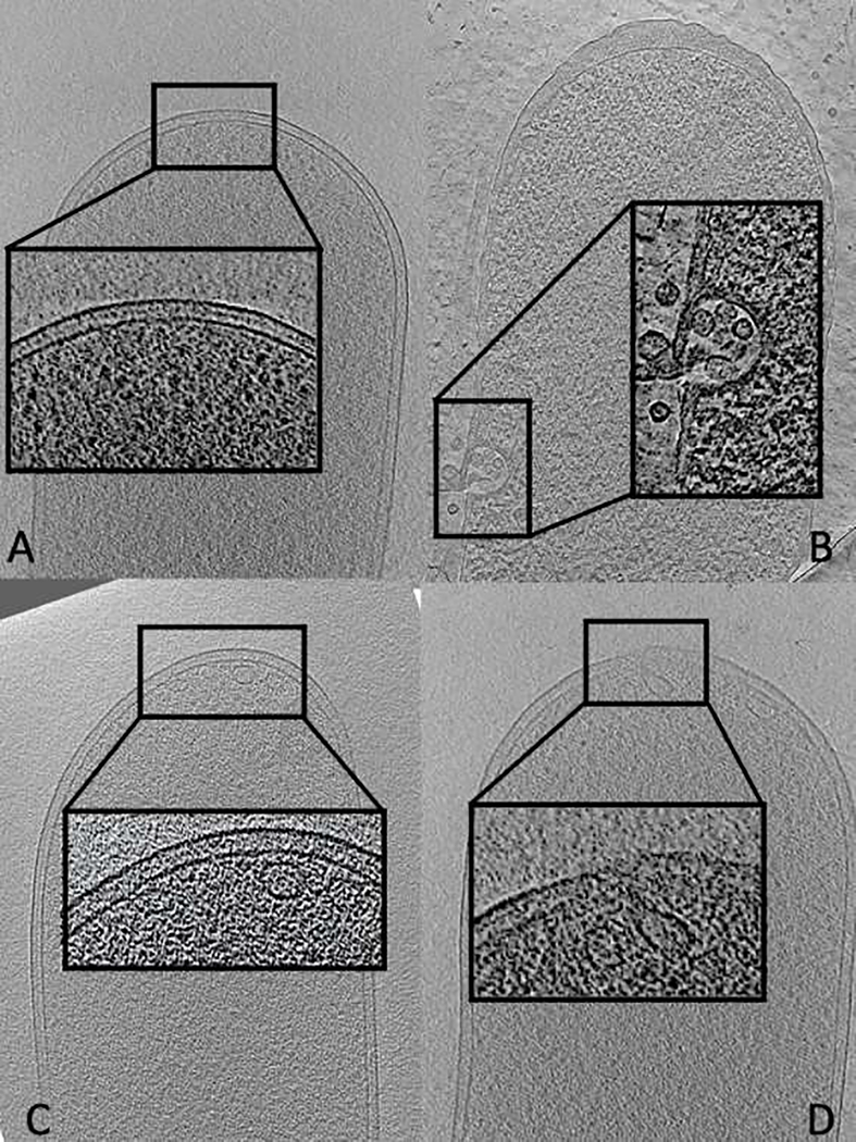
Figure 2:
Representative tomographic slices of cryo-section through E. coli cell treated with methylene blue and irradiated with 3J or 18J and treated with penicillin. A) Control group – no treatments. Note the presence of intact inner and outer membrane, such as cell wall B) Penicillin treated. Note the rupture of cell envelope with the presence of multiples vesicles C) aPDT 3J and D) aPDT 18J, presence of vesicles and buds. Zoom-in of cell envelope of each group.

