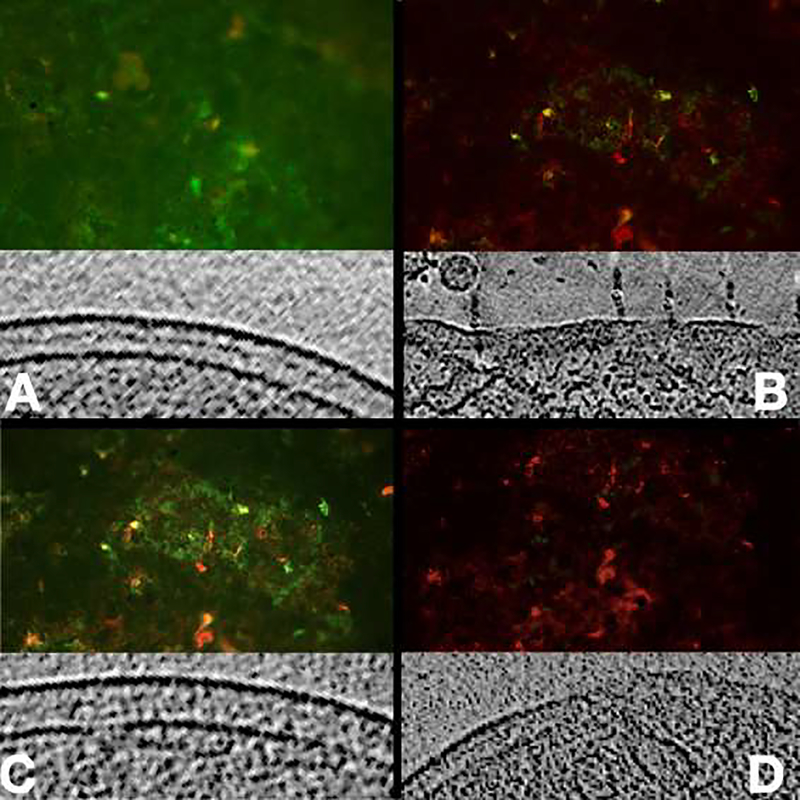
Figure 4:
Fluorescence images of biofilm on glass cover slides using live/dead stain and a representative tomographic slice of cryosection. A) Most of the bacteria are viable (green), note the intact envelope; B) Treatment with penicillin, significative damage to cell envelope; C) Sub-lethal dose of aPDT, some of the bacteria are dead, note the vesicles formation; D) Lethal aPDT dose, most of the bacteria are dead (red), vesicles and damage to cell envelope.

