Figure 10.

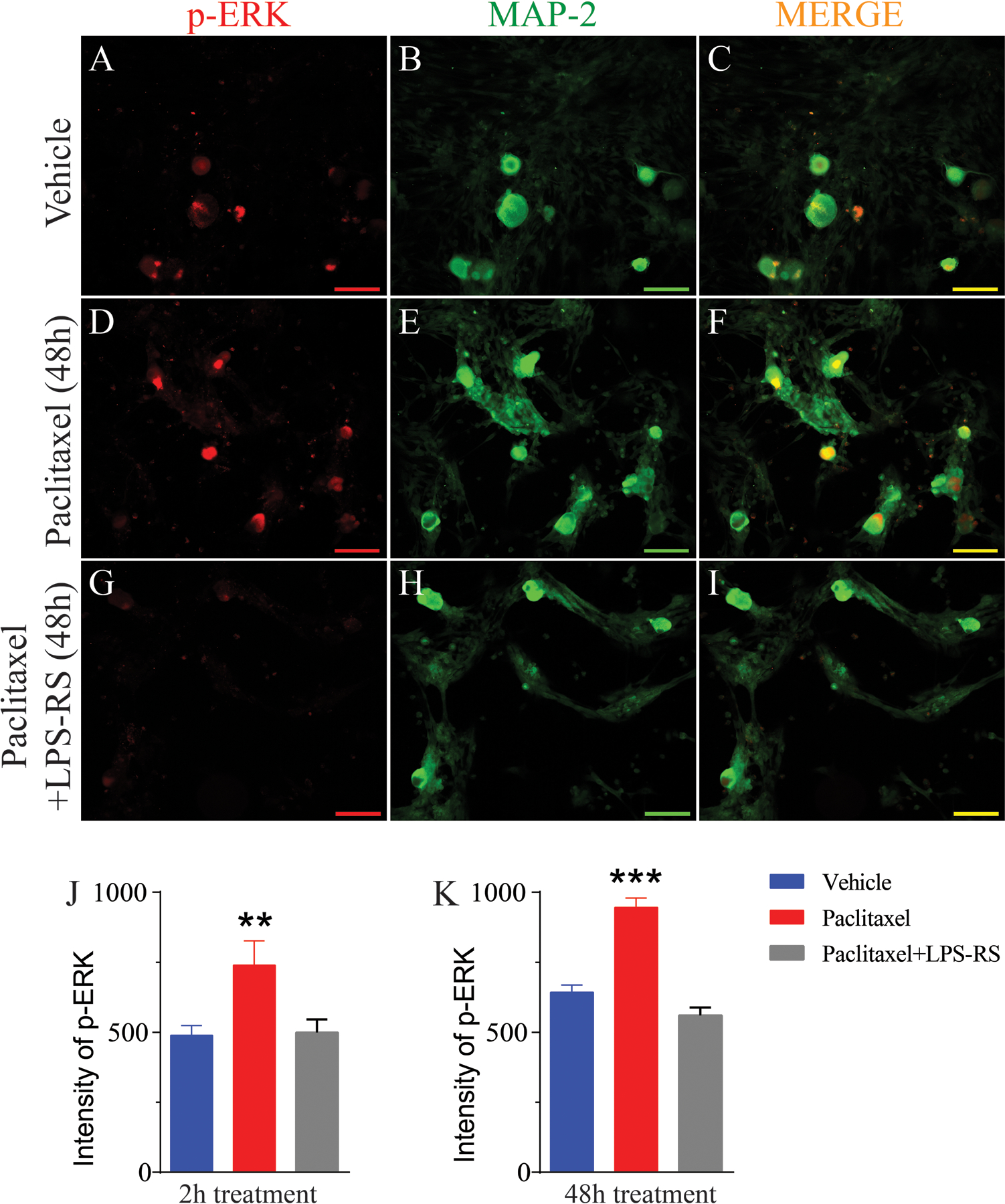
Double IHC revealed that pERK is increased in cultured human DRG neurons after incubation with paclitaxel. The representative immunohistochemical images in (A) and (D) show that the expression of pERK (red) in the DRG is low in vehicle-treated culture incubated for 48 h but that there is a marked increase in expression in culture treated with paclitaxel and incubated for 48 h. Upregulation of pERK were prevented by co-treatment with LPS-RS (G). Double immunohistochemical images shown in (C, F and I) indicate that pERK co-localized to MAP2 (B, E, and H, green)-positive cells (co-localization indicated by yellow). The bar graph in (J and K) shows that a significant increased fluorescence intensity of pERK at both 2 (n=10 neurons/group) and 48 h after paclitaxel treatment compared to vehicle-treated cultures and the upregulations were prevented by co-treatment with LPS-RS. Scale bar= 100 μm. ** = p <0.01, *** = P < 0.001.
