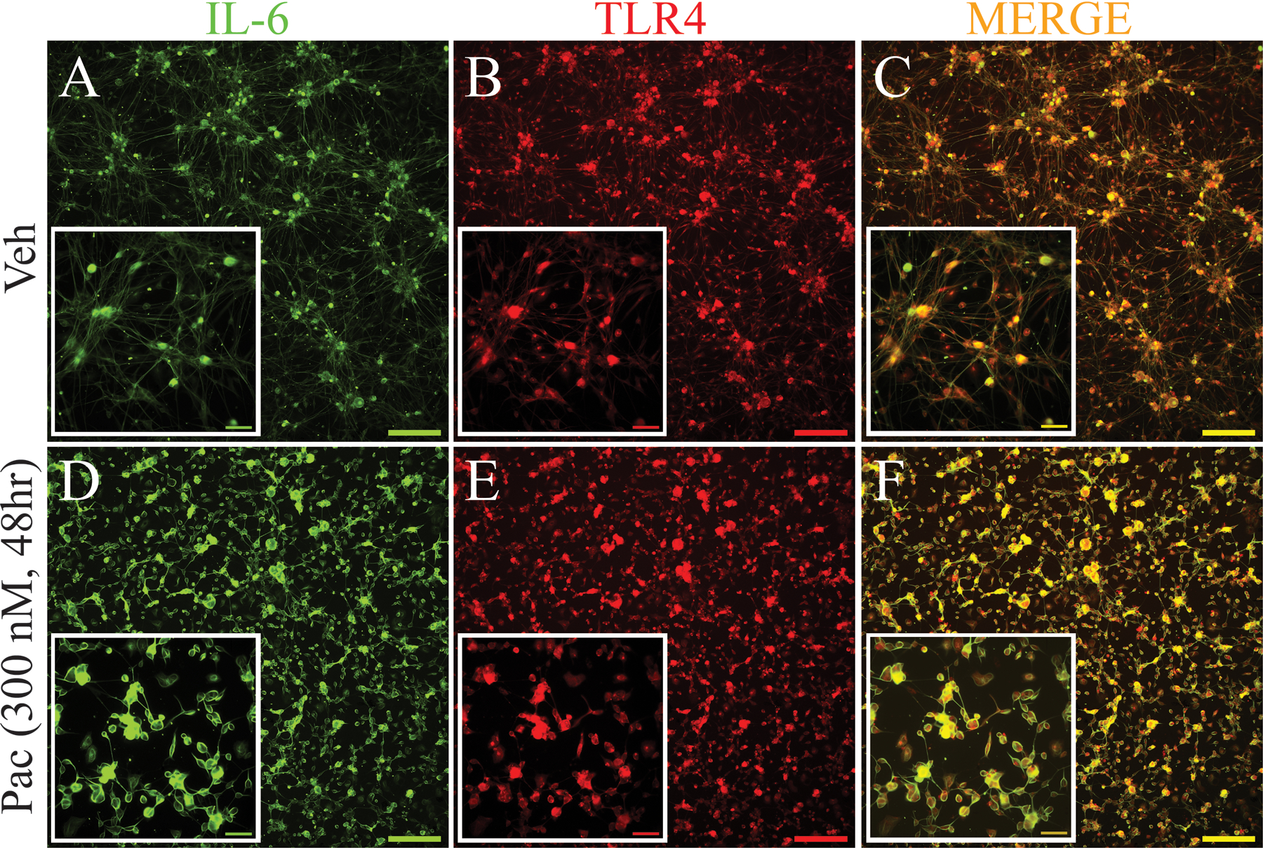
Figure 3.

Immunohistochemical stains in (A) and (D) show significant upregulation of IL-6 (green) in primary DRG culture at 48 h after incubation with paclitaxel compared with vehicle-treated culture. The immunohistochemical stains in B and E show significant upregulation of TLR4 (red) in DRG neurons in primary DRG culture at 48 h after incubation with paclitaxel compared with vehicle-treated culture. Double immunohistochemical images in (C) and (F) show that IL-6 (green) colocalized with TLR4 (red) in neurons in both vehicle and paclitaxel conditions. (C and F, colocalization indicated by yellow). Scale bar=200 μm and 100 μm in inserted pictures.
