Figure 5.

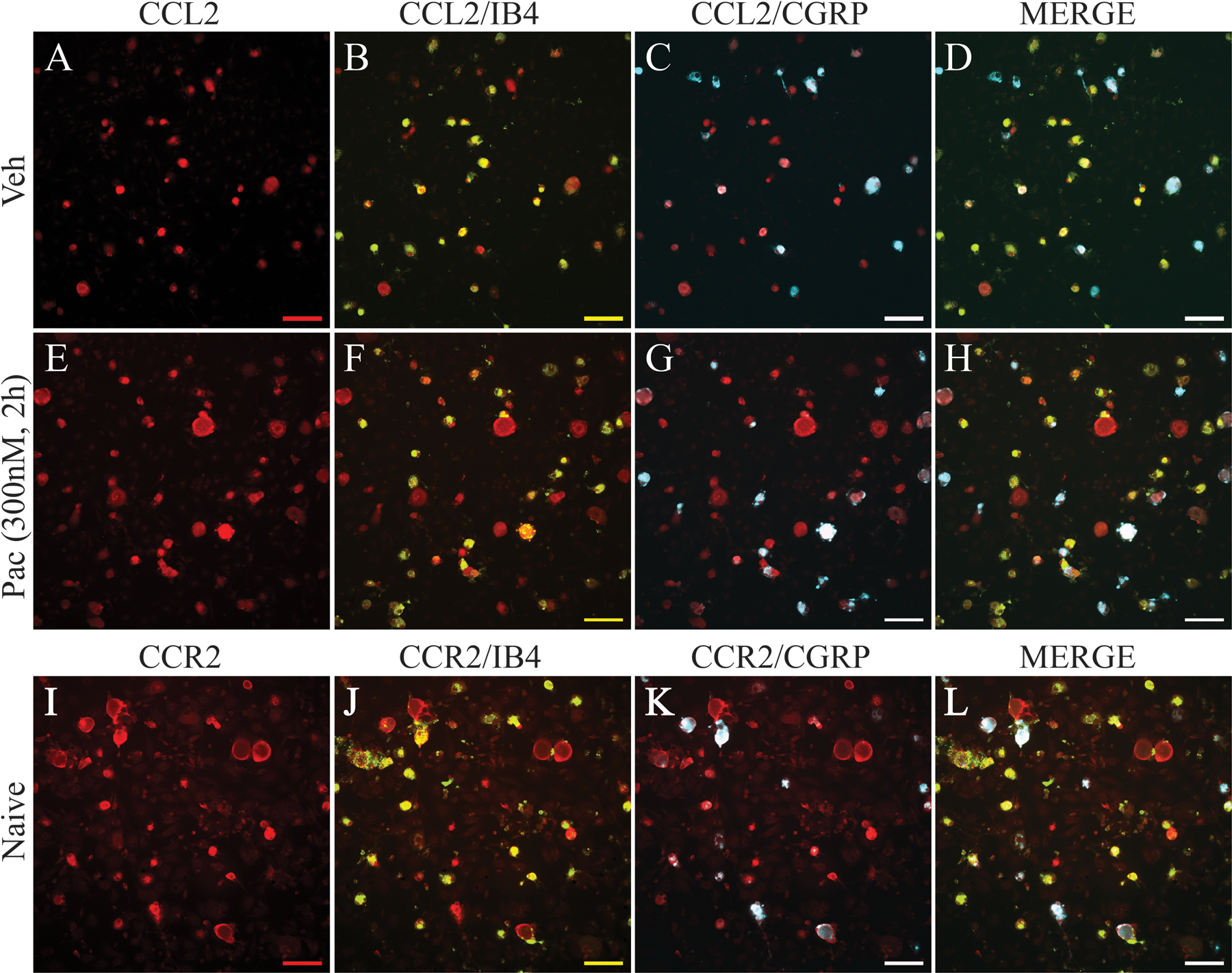
Immunostaining of primary DRG culture demonstrates that expression of CCL2 (red) is upregulated in neurons in paclitaxel-treated culture (E-H) compared with neurons in vehicle-treated culture (A-D) at 2 h after paclitaxel incubation. Images (B) and (F) show that CCL2 is co-localized to IB4-positive neurons (green, double label in yellow), and images (C) and (G) show that CCL2 is co-localized to CGRP-positive neurons (blue, double label in white) in both vehicle and paclitaxel conditions. Images (D) and (H) show CCL2, IB4, and CGRP labels merged. Immunostaining of naïve primary DRG culture (I-L) shows that CCR2 (red) strongly co-localized with IB4-positive neurons (J, green, double label in yellow) and to a lesser extent with CGRP-positive neurons (K, blue, double label in white). Images (L) shows CCR2, IB4, and CGRP labels merged. Scale bar=100 μm.
