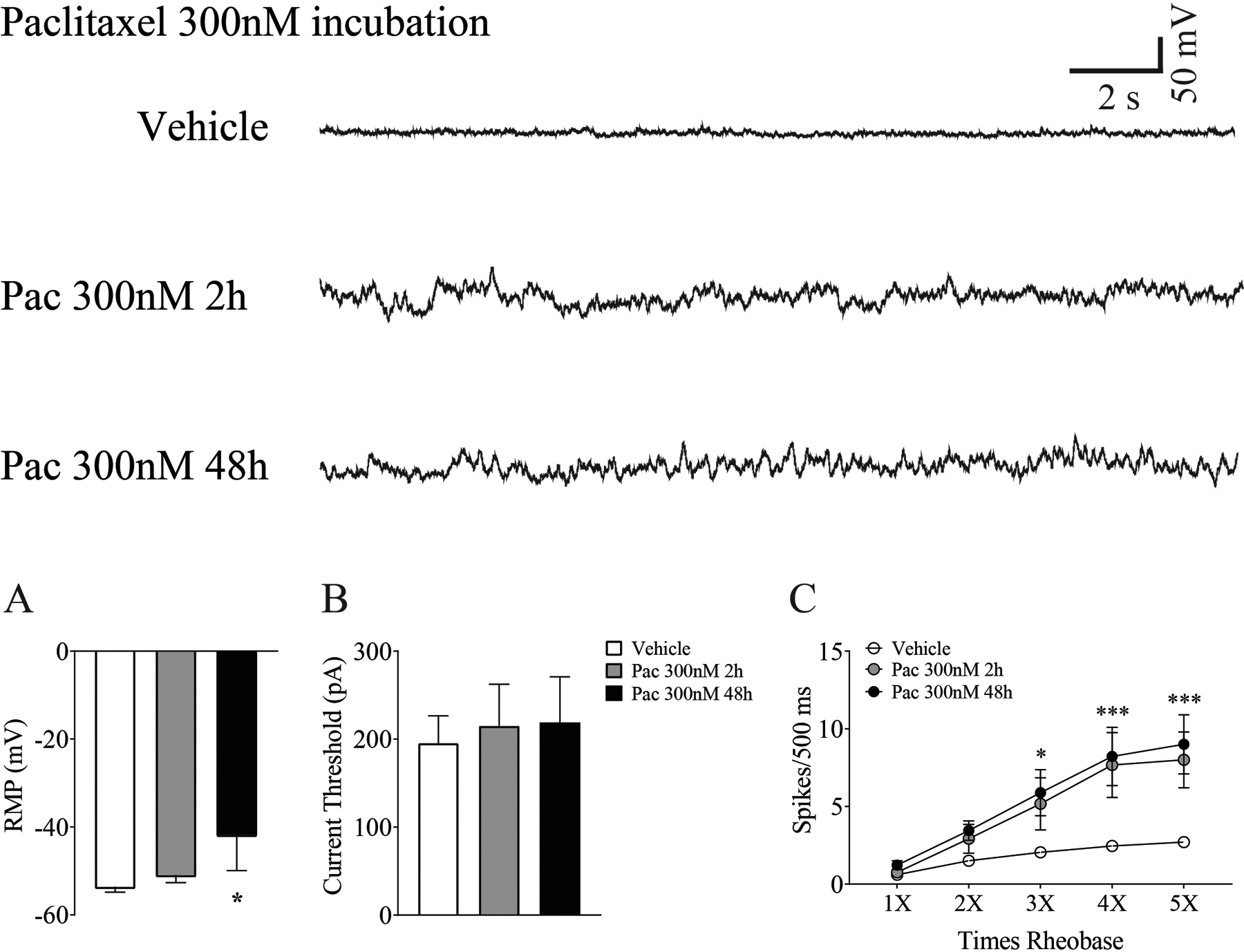
Figure 7.

Whole cell patch clamp recordings reveal paclitaxel-induced changes in DRG neurons in vitro. Depolarizing spontaneous fluctuations (DSFs) in membrane potential were seen after 2h and 48 h of paclitaxel incubation as presented in the traces at the top of the figure. DSFs were accompanied by a significant decrease in mean membrane potential (A), but no change in current threshold was observed at either time point (B) (n=15–25 cells/group). As summarized in C, neurons from cultures incubated with paclitaxel showed more evoked action potentials at 3 −5X rheobase compared to those incubated with vehicle (n=10/group/current strength). *p <0.05, ***p < 0.001.
