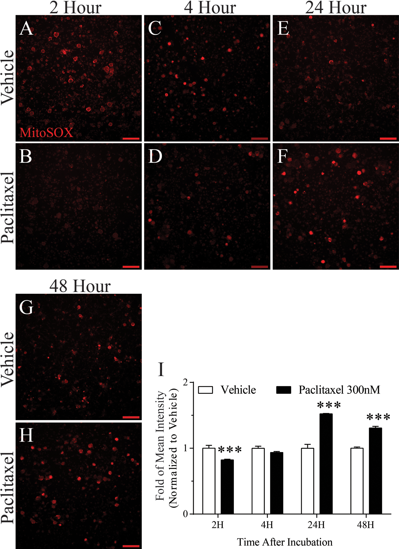
Figure 9.

Effects of paclitaxel-induced mitochondrial superoxide. Mitochondrial superoxide production in live DRG neurons was measured by fluorescence microscopy using MitoSOX Red dye. Representative fluorescence images show MitoSOX Red fluorescence at 2, 4, 24, and 48 h after vehicle or paclitaxel incubation (n=5/group/time point). Fluorescence intensity summaries in bar graph I, show that relative to vehicle, mitochondrial superoxide was lower after 2 h paclitaxel incubation and unchanged after 4 h. In contrast, mitochondrial superoxide was substantially higher in DRG neurons incubated with paclitaxel for 24 h or 48 h. ***p < 0.001, Scale bar 100 μm.
