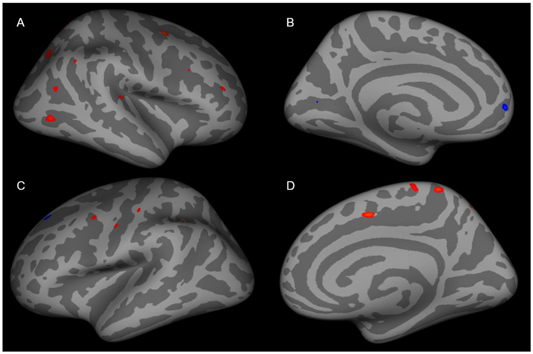
Figure 2: Cortical thickness in anxious adolescents with core depressive symptom burdens that are greater than the median core depression symptom score compared to those with fewer than the median core depression symptom score.

(A) Cortical thickness was increased in the caudal middle frontal, lateral occipital, rostral middle frontal, superior parietal, superior temporal and inferior parietal regions in the right lateral cortex. (B) Cortical thickness was decreased in the pericalcarine and superior frontal regions. In the left lateral cortex (C) cortical thickness was increased in the precentral, postcentral, supramarginal and superior parietal regions and decreased was observed in the superior frontal region. (D) Cortical thickness was increased in superior frontal, precuneus and paracentral regions.
