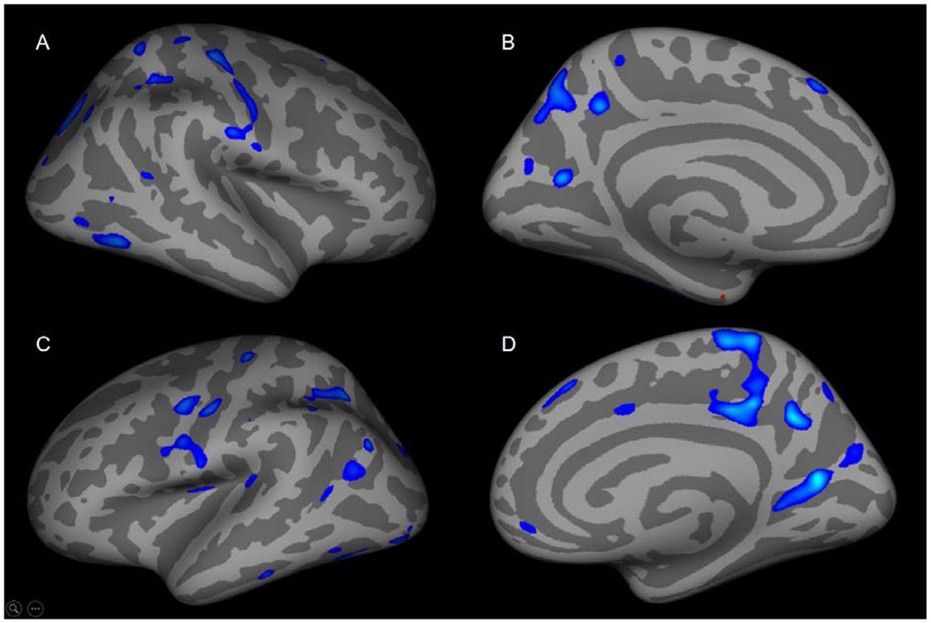
Figure 3: Age-cortical thickness correlations are decreased in anxious adolescents with greater core depressive symptoms.

In anxious adolescents with greater depressive symptoms, cortical thickness-age correlations were decreased in (A) inferior parietal, postcentral, lateral occipital, mid-temporal, superior frontal, inferior temporal, superior parietal and supramarginal regions. (B) Cortical thickness-age correlations were decreased in the precuneus, cuneus, superior frontal regions and was increased in the entorhinal region. (C) Cortical thickness was decreased in the superior parietal, pre- and postcentral, inferior parietal, supramarginal, inferior/superior temporal, lateral occipital, insula, superior temporal regions. (D) Cortical thickness-age relationships were decreased in the medial orbital frontal, posterior cingulate, precuneus, cuneus and superior frontal.
