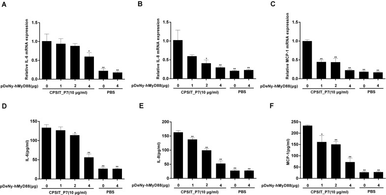
FIGURE 7.
CPSIT_P7 induced the expression of IL-6, IL-8, and MCP-1 in THP-1 cells via MyD88. THP-1 cells were transfected with 1, 2, or 4 μg of dominant negative plasmid encoding MyD88 (pDeNy-hMyD88) or empty vector (pDeNy-mcs) as a control for 48 h and were treated with CPSIT_P7 (10.0 μg/ml, 24 h) to detect the levels of IL-6, IL-8, and MCP-1 mRNA by RT-qPCR (A–C), or were treated with 10 μg/ml CPSIT_P7 for 36 h to detect the levels of IL-6, IL-8, and MCP-1 protein in the cellular supernatants by ELISA (D–F). The results shown are representative of three independent experiments. Data are means ± SD. *P < 0.05, **P < 0.01 vs. control group (10 μg/ml CPSIT_P7, 0 μg pDeNy-hMyD88).

