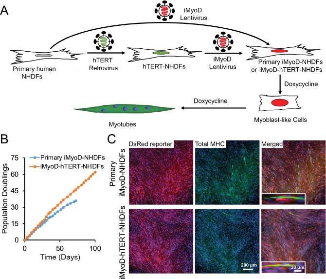
Figure 1.
Generation of primary iMyoD-NHDFs and iMyoD-hTERT-NHDFs and their myogenic transdifferentiation via induced MyoD expression. (A) Illustration of the procedures to generate these cells through viral transductions and to induce MyoD expression with DOX. (B) The extensive expansion capacity of iMyoD-hTERT-NHDFs endowed by hTERT as indicated by an almost unchanged growth rate at high population doubling values. (C) Representative images showing that both cell types expressed the DsRed reporter for iMyoD (red) and cells positive for total MHC (green) were present after 3-day DOX induction. The total MHC was stained with the MF20 antibody, and nuclei were stained with Hoechst 33342 (blue).

