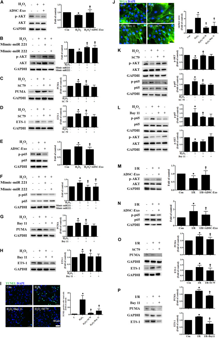
FIGURE 5.
ADSC-Exo reduced apoptosis and hypertrophy in H2O2-treated H9c2 cells through the AKT/NFκB pathway. Cardiomyocytes were pretreated with or without ADSC-Exos (2 μg/mL) for 4 h and then exposed to 50 μM H2O2 for 48 h. (A) The effect on AKT was examined in H2O2-treated H9c2 cells. H2O2 reduced the phosphorylation of AKT when compared to that in control cells, while ADSC-Exo increased AKT expression, as indicated by Western blot analysis. (B) Transfection of miR-221/222 mimics significantly ameliorated H2O2-reduced AKT phosphorylation. (C,D) Pretreatment with SC79 (10 μM) for 1 h reduced H2O2-induced PUMA and ETS-1 expression in H9c2 cells. (E) The expression of NFκB in H2O2-treated H9c2 cells was examined by Western blot analysis. H2O2 increased the phosphorylation of NFκB p65 compared to that in control cells, while ADSC-Exo reduced the expression of p-p65. (F) Transfection of the miR-221/222 mimics significantly reduced the increased phosphorylation of p65 by H2O2. (G,H) Pretreatment with an NFκB inhibitor (Bay 11, 2.5 μM) for 1 h reduced H2O2-induced PUMA and ETS-1 expression in H9c2 cells. (I,J) Pretreatment with SC79 or with Bay 11 for 1 h decreased apoptosis and hypertrophy in H2O2-treated cardiomyocytes, as detected by TUNEL assay and WGA staining, respectively. Quantitative analysis of TUNEL-positive cells or cell surface area is shown. (K,L) SC79 reduced the phosphorylation of NFκB, while Bay 11 did not affect AKT phosphorylation. (M,N) I/R treatment significantly decreased p-AKT and increased p-p65 compared to that in control mice, while ADSC-Exo reversed these effects in vivo. (O,P) Mice treated with SC79 (10 mg/kg) or with Bay 11 (80 mg/kg) exhibited reduced expression of PUMA and ETS-1 in I/R-treated mice. Data are expressed as the mean ± SD (n = 3). *P < 0.05 vs. control; †P < 0.05 vs. H2O2.

