
Figure 5. Deficiency of endogenous mouse IFITMs enhances SARS‐CoV‐2 infection.
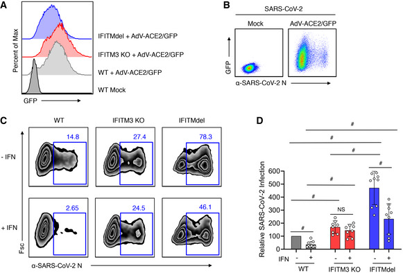
- WT, IFITM3 KO, or IFITMdel MEFs were transduced with adenovirus expressing human ACE2 and GFP (AdV‐ACE2/GFP) at an MOI of 100 for 48 h. Flow cytometry histograms show expression of GFP indicating near‐complete transduction in each MEF line.
- Flow cytometry plots of WT MEF cells with or without transduction with AdV‐ACE2/GFP for 48 h followed by infection with SARS‐CoV‐2 (MOI 1) for 24 h demonstrating that SARS‐CoV‐2 infection of MEFs can occur when transduced with AdV‐ACE2‐GFP.
- MEF cells transduced with AdV‐ACE2/GFP as in (B) were mock treated (−IFN) or treated with mouse IFNβ (+IFN) for 24 h followed by infection with SARS‐CoV‐2 (MOI 1) for 24 h. Representative example flow cytometry plots measuring percent infection are shown for each cell line and treatment condition.
- Graph depicts normalized infection percentage measurements from two experiments with cells infected as in (C) and one experiment with MOI 0.1. Bars represent averages with individual data points shown as circles. Error bars represent SD. # P < 0.05 compared to vector control by ANOVA followed by Tukey’s multiple comparisons test.
Source data are available online for this figure.